PHTY100 Lecture Notes - Lecture 7: Popliteus Muscle, Abductor Hallucis Muscle, Sesamoid Bone


ANAT LA week 7 Muscles of the Leg and Foot
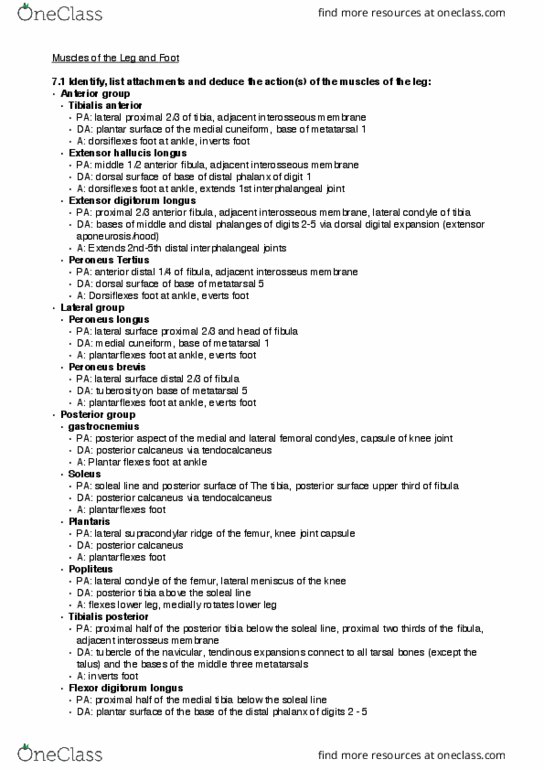
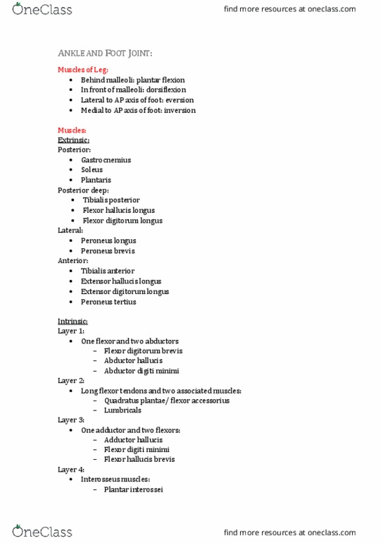
- Muscles of the leg – by compartment
- Additional foot features:
oRetinacula
oSynovial sheaths
oDorsal digital expansion
oPlantar aponeurosis
- Foot muscles – by layer
Muscles of the Leg (knee to ankle)
- We will group today by compartment
oPosterior
oAnterior
oLateral
- Fascia separates the superficial and deep posterior compartments
- Many two-joint muscles knee and ankle, ankle and tarsal joints and beyond
oWill need to consider actions at both joints
oWhen a muscle crosses two joints, action happens at the most distal joint
first
- Location of tendon at the ankle is a quick reliable way to begin determining action
oBehind the malleoli – performs plantarflexion
oIn front of the malleoli – performs dorsiflexion
oLateral to anterior-posterior axis of foot – performs eversion
oMedial to anterior-posterior axis of foot – performs inversion
Posterior group – superficial
- Three muscles:
oGastrocnemius (a) – big muscle
Two heads – medial and lateral head
Comes off medial and lateral condyles, crossing the knee
Technically can contribute to knee flexion but doesn’t have a great
line of pull for it
Sits superficial on top of soleus
oSoleus (b)
Doesn’t cross the knee
oPlantaris (c)
Vestigial muscle evolving out of them – some people don’t have
plantaris
Short muscle belly that becomes a long tendon
Insertion differs from person to person – either inserts beside
calcaneus or into calcaneus
- Note: gastrocnemius and soleus can be referred to as triceps surae they merge
with fascia to form Achilles tendon act together to plantarflex, postural muscles
find more resources at oneclass.com
find more resources at oneclass.com

Posterior group – deep
- Three muscles
oTibialis posterior
oFlexor hallucis longus (hallucis =
big toe)
Long muscle that flexes
big toe
oFlexor digitorum longus
Long muscle that flexes
toes
- When finding these in labs, Tom Dick
and Very Naughty Harry
oTom = Tibialis posterior
oDick = flexor digitorum longus
oVery = vein
oNaughty = nerve
oHarry = Flecor hallucis longus
- Popliteus is very deep and crosses back of knee but isn’t technically part of posterior
group because it doesn’t go down into foot
find more resources at oneclass.com
find more resources at oneclass.com

Lateral Group
- Two muscles
oPeroneus longus (also called fibularis longus)
Top 1/3
oPeroneus brevis (also called fibularis brevis)
- Both pull fibula
- Wrap around posterior to lateral malleolus
- Good at eversion and contribute extra for plantarflexion
Anterior Group
- Four muscles
oTibialis anterior
oExtensor hallucis longus
Extends big toe
oExtensor digitorum longus
Comes down as one body and splits into multiple tendons that go to
the four toes
oPeroneus tertius
- All dorsiflex the ankle
Tendons of anterior and lateral groups
find more resources at oneclass.com
find more resources at oneclass.com
Document Summary
Anat la week 7 muscles of the leg and foot. Muscles of the leg by compartment. Additional foot features: retinacula, synovial sheaths, dorsal digital expansion, plantar aponeurosis. We will group today by compartment: posterior, anterior, lateral. Fascia separates the superficial and deep posterior compartments. Many two-joint muscles knee and ankle, ankle and tarsal joints and beyond: will need to consider actions at both joints, when a muscle crosses two joints, action happens at the most distal joint first. Three muscles: gastrocnemius (a) big muscle. Comes off medial and lateral condyles, crossing the knee. Technically can contribute to knee flexion but doesn"t have a great line of pull for it. Sits superficial on top of soleus: soleus (b) Doesn"t cross the knee: plantaris (c) Vestigial muscle evolving out of them some people don"t have plantaris. Short muscle belly that becomes a long tendon. Insertion differs from person to person either inserts beside calcaneus or into calcaneus.