PHTY100 Lecture Notes - Lecture 11: Pubic Symphysis, Pelvic Brim, Pelvic Inlet

Document Summary
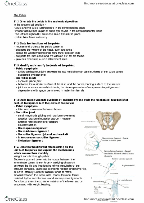
Pubic symphysis: fibro cartilaginous joint between two medial symphyseal surfaces of pubic bones, no movement between bones. Sacroiliac joint: synovial, plane, between auricular surface of ilium and corresponding surface of sacrum, develop a series of complementary ridges and depressions with age, movements: small magnitude gliding and rotation movements, ligaments: Pelvis: pelvic inlet- pelvic brim, pubic crest- pectineal line, arcuate line, sacral alar and promontory, pelvic outlet- tip of coccyx, ischial tuberosity"s, inferior part of pubic symphysis. Pelvic floor (pelvic diaphragm): two skeletal muscles (voluntary, levator ani, coccygeus, functions: Act as sphincters for the passages through the pelvis. Perineum: superficial to pelvic floor, contains skeletal muscle and the external genitalia, urogenital triangle: Act as sphincters for the openings and assist in maintaining erection: anal triangle: Allows anal canal to expand during defecation. Around an ap axis- pelvic lift and drop: corresponding lateral flexion of the lumbar spine (contralateral with pelvic drop, corresponding hip abduction with pelvic drop.