PHTY306 Lecture 7: PHTY306 Week 7 LEC 1

Document Summary
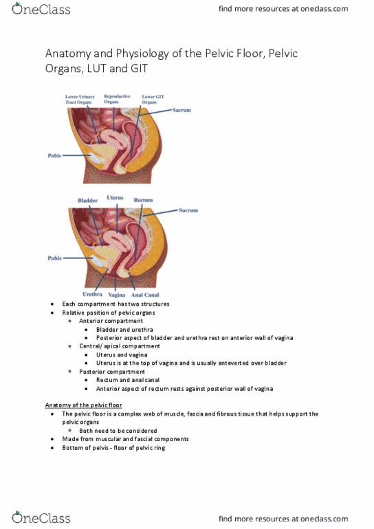
Relative position of pelvic organs to each other. The pelvic floor is a complex web of muscle, fascia and fibrous tissue that helps support the pelvic organs. : muscular components of the pf. 2 main layers: deep pelvic floor layer levator ani, coccygeus, superficial pelvic floor layer urogenital triangle, external anal sphincter. Deep pelvic floor (la: responsible for, forward pull of structures. Maintains anorectal angle - angle between rectum and anal canal : lift of pelvic structures ie levator . Lifts to support the pelvic organs, removing strain on the endopelvic fascia. Uterosacral ligaments: suspend lower aspect of uterus / cervix up and attach to sacral base, maintains uterus high in apical section of vagina, damage to uterosacral ligaments descent of uterus. Summary of the pf: the pelvic floor refers to the structures that support the pelvic organs . Therefore, when we talk about pelvic floor dysfunction, we could be talking about. Micturition 2 phases: storage phase, voiding phase.