ANAT20006 Lecture Notes - Lecture 6: Dura Mater, Pia Mater, Arachnoid Mater

12 Jun 2018
School
Department
Course
Professor

LECTURE 6
NEUROANATOMY
TODAY
•Anatomical orientation: the location of the nervous system
•Organisation the central nervous system
ANATOMICAL ORIENTATION
•(1) The nervous system (of vertebrates) comprises a
central division and a peripheral division.
•The CNS is encased within bony structures within the
body, and the spinal cord is within the vertebral column.
Emanating from these tissues is all the nerves that control the
body.
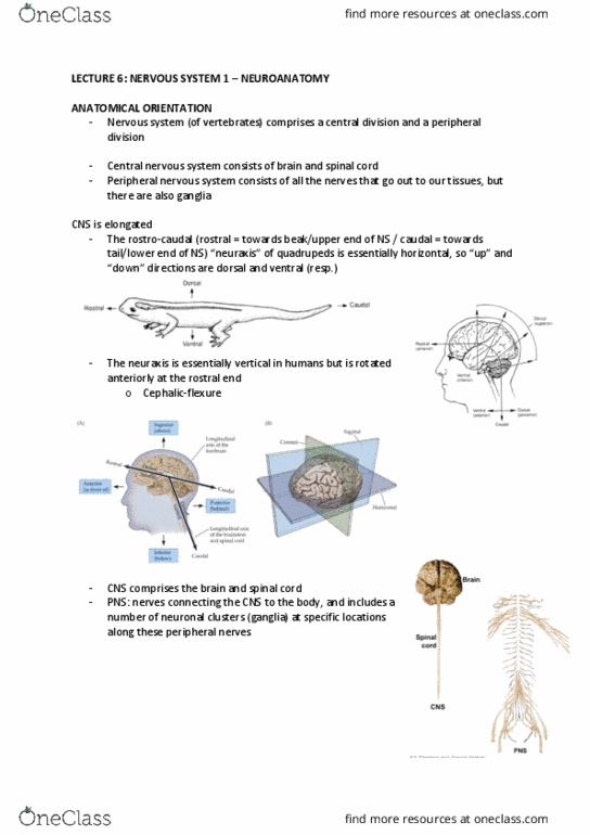
•(2) The rostro-caudal “neuraxis” of quadrupeds is essentially
horizontal, so “up” and “down” directions are dorsal
and ventral (resp.)
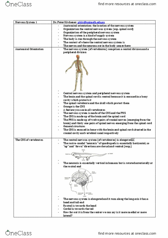
•The neuraxis is essentially vertical in humans but is
rotated anteriorally at the rostral end.
•Dorsal = superior, ventral = inferior (for brain).
•But spinal cord: dorsal = posterior, ventral = anterior.
•(3) orientation of planes throughout the NS. Coronal section
= end up with a front bit and back bit of the brain, if you do
it so you end up with left bit and right bit it is sagittal and
ending up with top and bottom is horizontal.
CNS & PNS
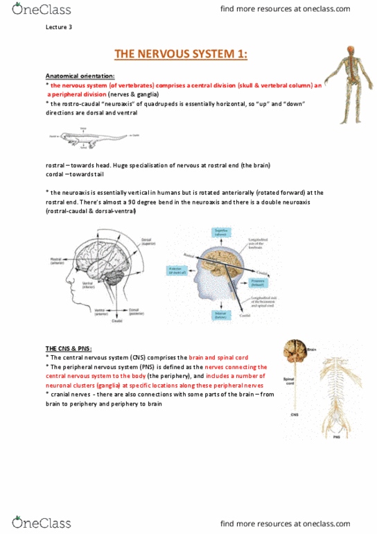
•(4) The central nervous system (CNS) comprises the brain and spinal cord.
Lecture 6 - Friday 4 August 2017
ANAT20006 - HUMAN STRUCTURE & FUNCTION

•The peripheral nervous system (PNS) is defined as the nerves connecting the central nervous
system to the body (the periphery), and includes a number of neuronal clusters (ganglia) at
specific locations along these peripheral nerves
•Brain made up of cerebrum, cerebellum, brain stem, diencephalon (which contains thalamus).
•Specialised functions in each hemisphere.
MENINGES
•(5) CNS important tissue for survival. Also is delicate.
•The Central Nervous System is wrapped in 3 membranes known as Meninges. They form a
protective coating. They are membranes that wrap
around the brain and spinal cord.
•Outermost layer = Dura mater (tough mother). It
is very fibrous, looks like a swimming cap.
Underneath it there is a subdural space which is
important bc blood vessels sometimes get
ruptured.
•(6) All three meninges: dura mater, arachnoid
mater and pia mater. The subarachnoid space is
filled with cerebrospinal fluid (CSF)
•Under the subdural space is the arachnoid
membrane/mater, under which is the
subarachnoid space, which is where CSF exists.
The brain is bathed in the fluid and this is where
CSF flows. The final layer is the
pia mater (delicate mother)
Delicate mother that covers the
brain. It is very thin and can’t be
separated from the hemispheres.
Pia mater dips down within
every convolution in the brain.
DURA MATER
•(7) The Dura Mater
•(8) the brain has a very specific
arrangement. The folds are called
gyri and the grooves between
them are called sulci.
This allows for a greater
surface area.
Lecture 6 - Friday 4 August 2017
ANAT20006 - HUMAN STRUCTURE & FUNCTION

CSF
•(9) CSF forms a protective cushioning
around the brain. Comes from the
ventricular system. CSF is inside the
brain (ventricles) and surrounds the
brain and spinal cord.
•Within the brain are 4 spaces called
ventricles. There is a lateral ventricle in
each hemisphere and 2 midline
structures. The ventricular system is
lined with epithelial cells. It contains
blood vessels and when blood flows into
it, some of the components are filtered
into these spaces with the CSF.
•The relationship between the ventricles
(where CSF is produced by the
specialised vascular tissue called choroid plexus) and the subarachnoid space diagram.
•CSF is formed within the ventricular system in the brain and
leaves the ventricular system to flow around the brain
in a sub arachnoid space.
MENINGES & CSF
•(10) CSF production is ongoing, as blood is always
moving. So to keep the balance there must always
be ongoing reabsorption of CSF. So CSF
reabsorption occurs via arachnoid vili, which are
part of the arachnoid membrane that protrudes up
into the blood vessels in the dura mater. So as CSF is
produced down there, it is reabsorbed at the same
rate via the arachnoid vili.
•(11) Hydrocephaly is when arachnoid vili don’t exist
and the liquid can’t be reabsorbed.
•(12) lateral view. The slimy stuff on the surface is the
arachnoid membrane. Can see it passing over the sulci,
not dipping in, so we know it is arachnoid.
BRAIN STRUCTURES
•(13) FPOT lobes. Each lobe isn’t arbitrarily defined.
There’s a bis sulcus sitting between the frontal and
parietal lobes. It is the central sulcus. It is the defining
separation separating the lobes.
•There is another sulcus between the parietal and
occipital lobe. There is a lateral fissure separating the
temporal lobe from the rest of the brain.
•(14) the brain is not homogenous in its function, different parts are involved in different things.
•However different functions tend to be clustered together.
•The gyrus next to the central sulcus in the frontal lobe is where the PMC is located, and most of
the other motor areas are toward the front of the frontal lobe. The big gyrus that sits behind the
central sulcus in the parietal lobe is the location of the PSSC. Most of the sensory info is directed
toward the back of the brain (Eg. Vision in occipital lobe, audition in temporal lobe).
Lecture 6 - Friday 4 August 2017
ANAT20006 - HUMAN STRUCTURE & FUNCTION
to brain
Document Summary
Today: anatomical orientation: the location of the nervous system, organisation the central nervous system. Anatomical orientation: (1) the nervous system (of vertebrates) comprises a central division and a peripheral division, the cns is encased within bony structures within the body, and the spinal cord is within the vertebral column. = end up with a front bit and back bit of the brain, if you do it so you end up with left bit and right bit it is sagittal and ending up with top and bottom is horizontal. Cns & pns: (4) the central nervous system (cns) comprises the brain and spinal cord. Also is delicate: the central nervous system is wrapped in 3 membranes known as meninges. They are membranes that wrap around the brain and spinal cord: outermost layer = dura mater (tough mother). It is very fibrous, looks like a swimming cap.