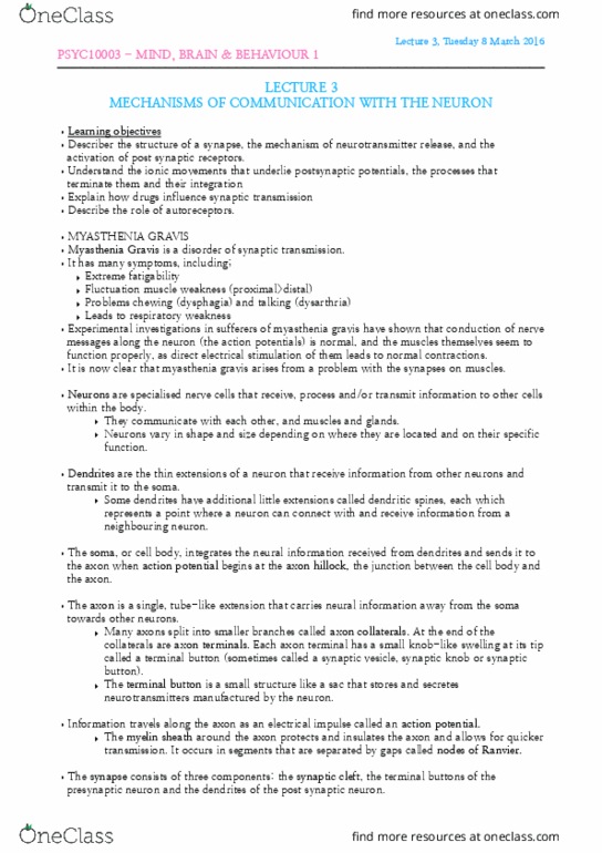
PSYC10003 Lecture Notes - Lecture 3: Myasthenia Gravis, Inhibitory Postsynaptic Potential, Synaptic Vesicle


3. The Synapse: Mechanisms of Communication between
neurons
Myasthenia Gravis – a disorder of synaptic transmission
• Myasthenia Gravis: grave muscle weakness
• Described by Thomas Willis in 1672
• First symptoms:
o Extreme fatigability
o Fluctuating muscle weakness
o Tends to affect proximal muscles (those of the head, neck and trunk) than distal muscles
(those of arms and legs)
o Dysphagia: problems chewing
o Dysarthria: problems talking
o Due to weakness of musculature of jaw and mouth, respectively
o In severe cases, respiratory distress: problems breathing
• Experimental investigations of sufferers shown that conduction of nerve messages along the
neuron (i.e. action potentials) is normal.
• Muscles themselves seem to function properly, as direct electrical stimulation leads to contractions
• Myasthenia gravis arises from a problem with the synapses on the muscles
The synapse — a means of communication between neurons
• Transmission of information within a neuron involves generation of action potential (AP)
• Begins at cell body (at junction between cell body and axon: axon hillock)
• AP proceeds along axon between the Nodes of Ranvier
• Once AP reaches terminal buttons how does it communicate with next neuron, despite not being
physically joined?
• Terminal buttons release a chemical message: neurotransmitter
• It diffuses across gap (synaptic cleft) between presynaptic terminal button (terminal button before
the synapse) and the dendrite or cell body of the postsynaptic membrane (membrane of the
neuron after the synapse)
• If neurotransmitter has an excitatory effect on the postsynaptic cell, then it will depolarise the
postsynaptic neuron and generate and AP
• Whole process repeated for next neuron in the circuit
• If neurotransmitter is inhibitory, postsynaptic cell will become hyperpolarised and therefore not fire
find more resources at oneclass.com
find more resources at oneclass.com

2
Structure of a synapse
3 types of synapses, defined on the basis of the places at which they occur
1. Axodendritic: the terminal button synapses with a dendrite of the postsynaptic neuron
2. Axosomatic: the terminal button synapses with the cell body (soma) of the postsynaptic
neuron
3. Axoaxonic: the terminal button synapses with the axon of the postsynaptic neuron
• Presynaptic membrane – the
membrane of the presynaptic
terminal button
• Postsynaptic membrane – the
membrane of the postsynaptic
neuron
• Dendritic spine – a ridge on the
dendrite of a postsynaptic neuron,
with which a terminal button from
a presynaptic neuron forms a
synapse
• Synaptic cleft – the tiny gap
between the presynaptic and
postsynaptic membrane
(approximately 20 nanometres
wide; a nanometre is a billionth of a
metre)
• Synaptic vesicles – tiny balloons
filled with neurotransmitter
molecules; found in the release zone of the terminal button
• Microtubules – long tubes that run down the axon and guide the transport of synaptic vesicles from
the soma to the axon terminal
• Release zone – part of the interior of the presynaptic membrane to which synaptic vesicles fuse in
order to release their neurotransmitter into the synaptic cleft
Release of a neurotransmitter – 1
• When AP is conducted down an axon (including all of
its branches), synaptic vesicles located just inside the terminal
buttons begin to move toward the release zone of the cell
membrane
• The vesicles are guided toward the cell membrane of
the presynaptic neuron by a group of protein structures (P)
Release of a neurotransmitter – 2
• Protein structures act like ropes, helping to pull the
vesicles towards the presynaptic membrane
find more resources at oneclass.com
find more resources at oneclass.com

3
Release of a neurotransmitter – 3
• An influx of calcium Ca2+ ions into the presynaptic neurons
o Induces fusion of membranes of synaptic vesicle and presynaptic cell
• Neurotransmitter molecules carried by synaptic vesicles then released into synaptic cleft
• Process occurs rapidly (few milliseconds)
Activation of receptors on postsynaptic neurons
• How do molecules of the
neurotransmitter released by terminal
buttons of presynaptic neuron
influence the postsynaptic cell?
• Neurotransmitter molecules diffuse
across fluid filled space of synaptic
cleft
• When they reach other side, attach to
specific binding sites of postsynaptic
receptors, located in the membrane of
postsynaptic cell (like a key to a lock)
• The neurotransmitter molecules open
neurotransmitter dependent ion channels in the postsynaptic cell
• These channels, once opened, permit the flow of specific ions into and out of the post synaptic
neuron
Neurotransmitters open ion channels in two different ways, direct and indirect
• Here we consider direct channel (simpler to understand):
o Involves receptors that are equipped with their own binding sites: ionotropic receptors
o When neurotransmitter molecule locks into the binding site, channel is opened allowing
ions to move in or out
find more resources at oneclass.com
find more resources at oneclass.com
Document Summary
The synapse: mechanisms of communication between neurons. It diffuses across gap (synaptic cleft) between presynaptic terminal button (terminal button before the synapse) and the dendrite or cell body of the postsynaptic membrane (membrane of the neuron after the synapse) If neurotransmitter has an excitatory effect on the postsynaptic cell, then it will depolarise the postsynaptic neuron and generate and ap: whole process repeated for next neuron in the circuit. If neurotransmitter is inhibitory, postsynaptic cell will become hyperpolarised and therefore not fire. When ap is conducted down an axon (including all of. Release of a neurotransmitter 1 its branches), synaptic vesicles located just inside the terminal buttons begin to move toward the release zone of the cell membrane. The vesicles are guided toward the cell membrane of the presynaptic neuron by a group of protein structures (p) Release of a neurotransmitter 2 vesicles towards the presynaptic membrane. Protein structures act like ropes, helping to pull the.