PSYC10003 Lecture Notes - Lecture 4: Arachnoid Mater, Pia Mater, Dura Mater

12 Jun 2018
School
Department
Course
Professor

Lecture 4, Wednesday 9 March 2016
PSYC10003 - MIND, BRAIN & BEHAVIOUR 1
LECTURE 4
STRUCTURE & FUNCTION OF THE HNS 1
STRUCTURE & FUNCTION OF THE HUMAN NERVOUS SYSTEM
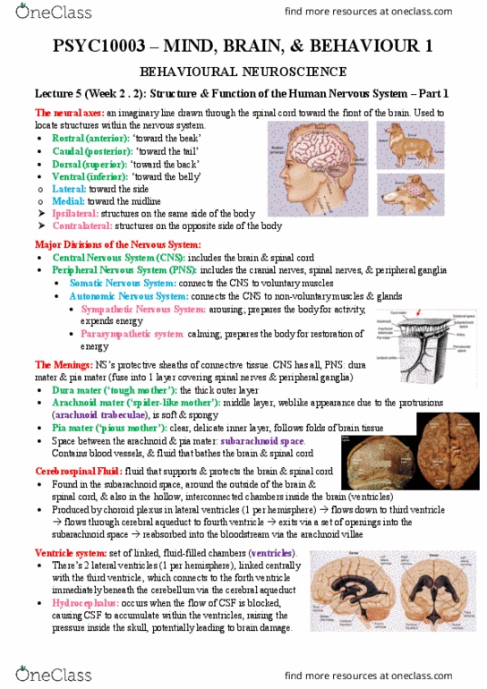
THE NEURAL AXES
•Rostral: ‘toward the beak’
•Caudal: ‘toward the tail’
•Dorsal: ‘toward the back’
•Ventral: ‘toward the belly’
•Because humans stand upright, their neuraxis bends.
•The top of the head is perpendicular to the back.
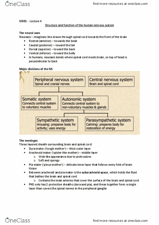
MENINGES
•The entire nervous system (CNS and PNS) is covered by a protective sheath of connective tissue.
•The protective sheaths around the brain and spinal cord are called the meninges (plural). In the
CNS there are three layers:
•1) Dura mater (tough mother): the thick outer layer
•2) Arachnoid mater (spider-like mother): the middle layer, which has a weblike appearance
due to the protrusions called arachnoid trabeculae, and is soft and spongy.
•3) Pia mater (pious mother): the delicate inner layer, which follows every fold of brain tissue.
•Lying between the arachnoid mater and the pia mater is the subarachnoid space, which holds the
fluid that bathes the brain and spinal cord, and which contains the main arteries that cover the
surface of the brain and spinal cord.
•The PNS has only two protective sheaths, the dura mater and the pia mater, and these fuse
together to form a single layer that covers the spinal nerves and peripheral ganglia.
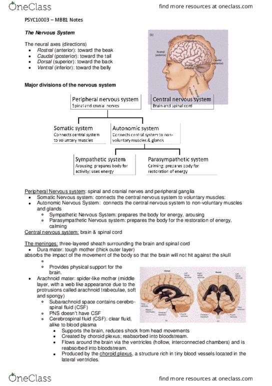
CEREBROSPINAL FLUID
•The brain is a bit like a jelly in a lunchbox. At an average weight of 1400 grams it needs to be
supported inside the bony cavity of the skull. This is achieved by bathing the brain in a protective
raft of fluid, called cerebrospinal fluid (CSF). The CSF supports the brain, and reduces its net
weight to about 80 grams.
•CSF is a clear fluid similar to blood plasma. It resides in the subarachnoid space around the
outside of the brain and spinal cord, and also fills the hollow, interconnected chambers inside the
brain known as the ventricles (remember that many early philosophers and physicians, from
Hippocrates through to Descartes, believed the ventricles were the seat of the mind).
•CSF is produced by the choroid plexus, a structure rich in tiny blood vessels located in the lateral
ventricles (there are two of these, one in each brain hemisphere). From the lateral ventricles CSF
flows down to the third ventricle, then through the cerebral aqueduct to the fourth ventricle. From
here it exits via a set of openings
into the subarachnoid space,
before being reabsorbed back
into the bloodstream via the
arachnoid villae.
THE VENTRICLES
•The ventricular system in the
brain consists of a set of linked,
fluid-filled chambers. The
lateral ventricles are located
within each hemisphere (one on
each side). These are linked
centrally with the third ventricle,
which is located in the midline

Lecture 4, Wednesday 9 March 2016
PSYC10003 - MIND, BRAIN & BEHAVIOUR 1
of the brain. A long tube called the cerebral aqueduct connect the third ventricle to the fourth
ventricle, which sits immediately beneath the cerebellum.
OBSTRUCTIVE HYDROCEPHALUS
•Occasionally the flow of CSF is blocked somewhere in its journey between the choroid plexus
within the lateral ventricle and the arachnoid villi within the subarachnoid space which channel it
back into the bloodstream.
•Such blockages cause a condition called hydrocephalus , in which CSF accumulates within the
ventricles because it is not reabsorbed into the bloodstream. This raises pressure inside the skull,
and can damage brain tissue and occlude arteries, leading to permanent (sometimes fatal) brain
damage.
•Hydrocephalus can be treated by inserting a ventriculo-peritoneal (VP) shunt. A hole is drilled in
the skull (under anaesthesia of course!) and a fine tube is inserted into one of the ventricles. The
tube runs beneath the skin, down into
the person’s abdominal cavity (the
peritoneum), from where it can be
reabsorbed into the bloodstream.
When pressure starts to increase in the
ventricles, a release valve in the tube
opens and the excess CSF is allowed to
flow out.
DEVELOPMENT OF THE CNS
•The nervous system begins to develop
around 18 days after conception. The
embryo begins as a plate of cells,
whose edges form ridges that curl
toward one another and fuse, forming a tube (the neural tube) that extends longitudinally from
rostral to caudal.
•At about 28 days after conception to neural tube has differentiated to form three interconnected
chambers. These chambers are destined to become the ventricles, and the surrounding tissue will
form the three main components of the adult brain: the forebrain, the midbrain and the
hindbrain. The tail connected to the hindbrain chamber will form the spinal cord.
•Later in development the tissue of the forebrain, midbrain and hindbrain differentiate to form the
precursors of the major structures present in the adult brain. The chamber of the forebrain divides
to form the two lateral ventricles and the third ventricle. The chamber inside the midbrain
narrows to form the cerebral aqueduct, and the chamber inside the hindbrain becomes the fourth
ventricle.
ANATOMICAL SUBDIVISIONS OF THE BRAIN
•All of the major structures of the brain can be associated with one of the three early precursors,
the forebrain, midbrain and hindbrain. As mentioned earlier each of the precursors consists of a
hollow chamber that
will eventually form
one of the ventricles.
In the fully formed
brain there are five
major subdivisions:
telencephalon,
diencephalon,
mesencephalon,
metencephalon and
myelencephalon (the
word cephalo means
Document Summary
Structure & function of the human nervous system. The neural axes: rostral: toward the beak", caudal: toward the tail", dorsal: toward the back", ventral: toward the belly", because humans stand upright, their neuraxis bends, the top of the head is perpendicular to the back. Meninges: the entire nervous system (cns and pns) is covered by a protective sheath of connective tissue, the protective sheaths around the brain and spinal cord are called the meninges (plural). Cerebrospinal fluid: the brain is a bit like a jelly in a lunchbox. At an average weight of 1400 grams it needs to be supported inside the bony cavity of the skull. This is achieved by bathing the brain in a protective raft of fluid, called cerebrospinal fluid (csf). The csf supports the brain, and reduces its net weight to about 80 grams: csf is a clear fluid similar to blood plasma.