IMED3002 Lecture Notes - Lecture 2: Cerebral Crus, Spinal Trigeminal Nucleus, Substantia Nigra

28 May 2020
School
Department
Course
Professor
Document Summary
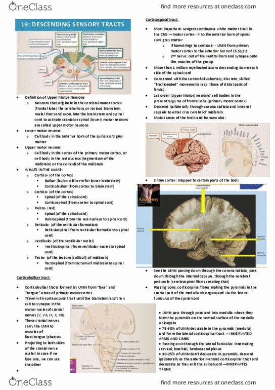
Clinical image: brownish structure: substantia nigra: basal portion: 2. Section: sagittal with medial view: brainstem: below the thalamus and hypothalamus, Looking at the left cerebral hemisphere anterior to the cerebellum and superior to the spinal cord: most superior part: cerebral peduncles (anterior part of the midbrain) Pons: medulla oblongata: inferior to the pons, between the pons and spinal cord. The clinical mri: t1 as the csf is not fluorescent, can see how the structures look similar on the. Four components of brainstem: tectum (posterior/dorsal on the back part of the brainstem): roof, ventricular system: part of this will run through it, tegmentum: middle portion, basal portion (anterior/ventral): Top picture: sagittal section looking at medial view. Anterior and posterior brainstem: has transverse strips on it- because these strips are going to the laterally to the cerebellum (pontine fibres going to the cerebellum) pons. Superior: connects the 2 cerebral superior cerebellar peduncles on the back of the pons.