MEDRADSC 2RA3 Lecture Notes - Lecture 3: Choroid Plexus, Septum Pellucidum, Interthalamic Adhesion

28 Jul 2019
School
Department
Course
Professor
Document Summary
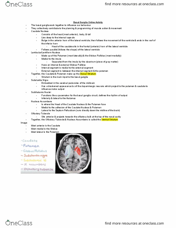
Lateral ventricles (x2): have 1 in each cerebral hemisphere. Most lateral ventricles; specifically the temporal horn of the lateral ventricle. Won"t ever see the whole lateral ventricle bc of its c shape. The 2 ventricles are separated by a membrane called the septum pellucidum. Considered to be a true midline structure in the brain (doesn"t deviate/change from patient to patient) If the partition is no longer midline, a midline shift has occurred & there is some pathology causing the partition to move. The junction of the body, occipital, & temporal horns of the lateral ventricles is called the trigone. Connected to the 3rd ventricle via the foramen monroe. Image is in the posterior aspect of the head bc seeing the cerebellum. Picking up the posterior horn of the lateral ventricle. Anterior horns of the lateral ventricle sits in the frontal lobes. Sinuses always look black; ventricles look black or white.