MEDRADSC 3L03 Lecture Notes - Lecture 6: Maxillary Sinus, Mandibular Symphysis, Foramen Magnum

10 Jun 2020
School
Department
Course
Professor
Document Summary
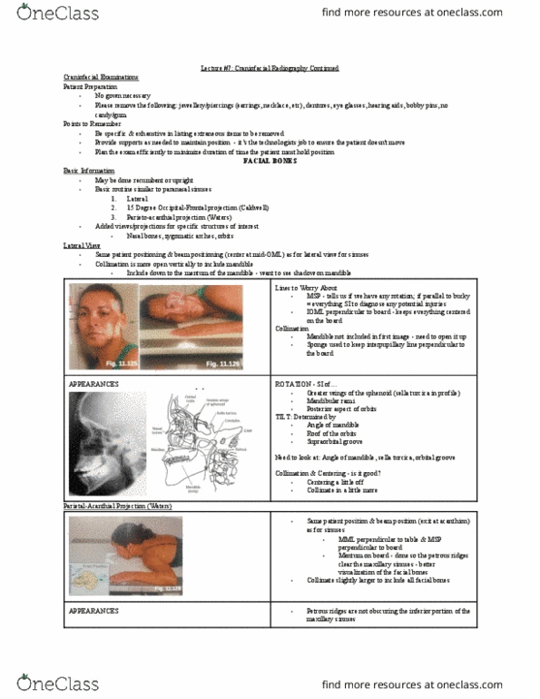
Challenges of craniofacial image: round, spherical structure. When they lay their head flat - most likely not flat. Slight rotation shows - will have to repeat. Area that is superimposed on other structures. Images: all show the petrous ridge in different spots. All facial structures are moving down & below the skull (bc of the caudal angle) Petrous - make a v out from the foramen magnum. Bc they are in the lower part, we have angled down in a. Pa position - when we line up a pa we line up the ioml perpendicular to the ir, then angle down - causes ridges to go below the orbits. When aligned, the petrous fill the orbits completely (when no angle) One of the views where we can see the ethmoid sinuses (medial to the orbits) Petrous are down at the very bottom of image; superior to symphysis menti. Clear view of maxillary sinuses (when done right)