ANAT 215 Lecture Notes - Lecture 9: Medulla Oblongata, Lateral Geniculate Nucleus, Anterior Chamber Of Eyeball

12 Apr 2016
School
Department
Course
Professor
Document Summary
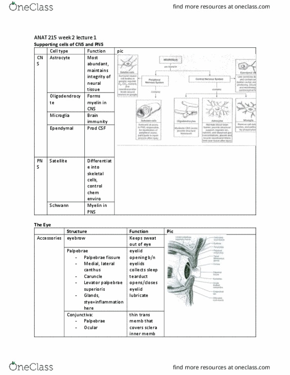
Extrinsic eye muscles: there are 6 extrinsic muscles of the eyes which are responsible for the movement of the eyeballs in cardinal direcions, up, down, let and right. Anterior and posterior poles: anterior cavity. Drains at juncion of cornea and sclera (canal of schlemm: posterior cavity. Not replaced layers of the eye (tunics): ibrous, vascular, sensory (reina) ibrous tunic, sclera. Cells receive nutrients from tears vascular tunic: choroid. Delivers oxygen and nutrients to inner layer (reina) Ends at ciliary body anteriorly: ciliary body: Begins at juncion of cornea and sclera. Diameter of pupil controlled by pupillary muscles. Pupil, pupillary sphincter and dilaing muscles (intrinsic eye muscles) sensory tunic (reina): pigmented epithelium layer, neural layer: Photoreceptors rods: no discriminaion of colour, 1 type, Less sharp, fuzzier images, very sensiive to light cones: discriminates between colour, 3 types (red, blue, green), sharp, clear.