PHGY 214 Lecture Notes - Lecture 4: Ventricular Fibrillation, Tachycardia, Qrs Complex

24 Jan 2017
School
Department
Course
Professor
Document Summary
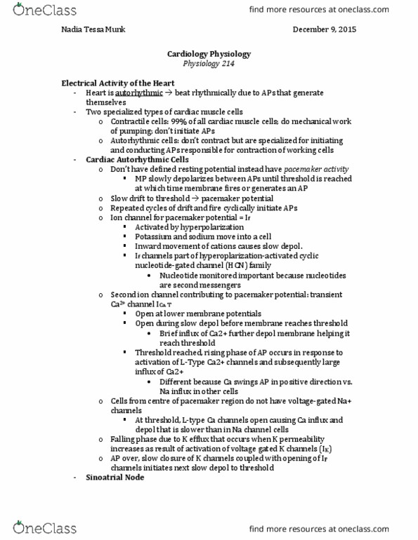
Electrical conduction system of the heart (action potential propagation) Lecture objectives: describe electrical conduction through the heart. Electrical conduction in myocardial cells depends on gap junction. Depolarizations of autorhythmic cells rapidly spread to adjacent contractile cells through gap junctions. Normal rate of action potential discharge in autorhythmic tissues. The autorhythmic cells have short and sharp action potentials with longer refractory period. (phase 4 of purkinje fibres pacemaker activity at the bottom) The contractile cells have a longer action potentials and a shorter refractory period. Electrical activity that originates from the sa node is propagated to the rest of the heart. Sa node right & left atrial muscle and intermodal conducting fibres. Internodal conducting fibres av node (slow) his bundle purkinje fibre conducting system ventricular muscle. [type here: distinguish/correlate ecg with cardiac action potentials. The voltage difference between excited and non-excited parts of the myocardium can be recorded on the body surface as electrocardiogram (ecg) or elektrokardiogram (ekg, german)