BIOL 1030H Lecture Notes - Lecture 13: Alpha Helix, Peptide, Hemoglobin

13 May 2018
School
Department
Course
Professor

Supplement
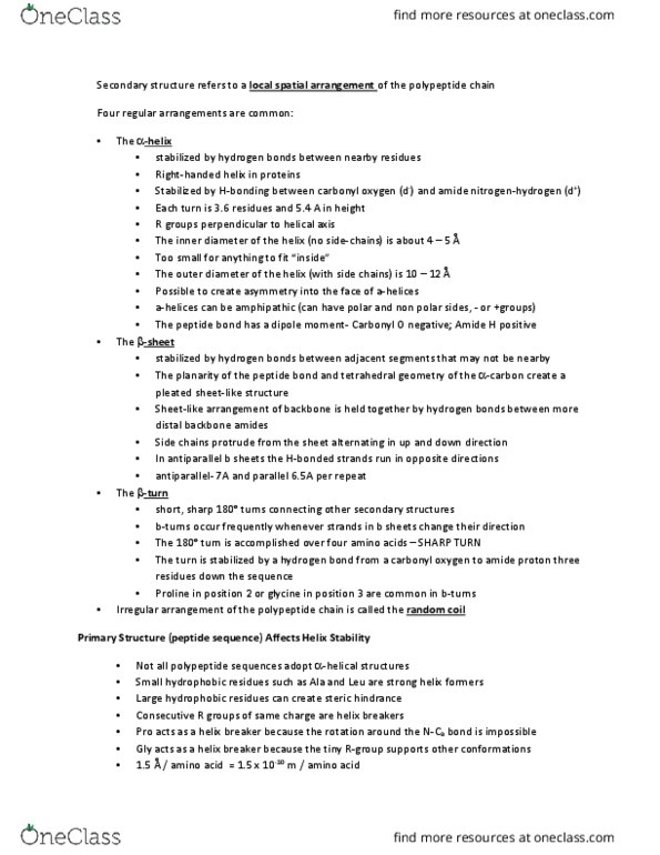
Secondary structure
●Alpha helix: polypeptide chain is twisted tightly in a right-handed coil
●Stabilized by hydrogen bonds between carbonyl group and the amide group four residues
ahead
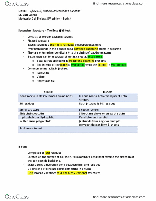
Beta Sheet
●Adjacent strands can run in the same direction (parallel), or in opposite directions
(antiparallel), as shown here.
●Hydrogen bonds form between carbonyl groups in one polypeptide and amide groups in a
different part of the polypeptide.
●The polypeptide folds back and forth on itself, forming a pleated sheet
Tertiary Structure
●Tertiary structure: three-dimensional shape, usually made of several secondary structure
elements
●Distribution of hydrophilic and hydrophobic R groups and chemical bonds/interactions
that form between the R groups
Quaternary Structure
●This enzyme consists of two identical polypeptide subunits, shown in light green and
dark green.
●Hemoglobin is made up of four subunits: two copies of the polypeptide depicted in
magenta and two copies of the polypeptide depicted in blue
Document Summary
Alpha helix: polypeptide chain is twisted tightly in a right-handed coil. Stabilized by hydrogen bonds between carbonyl group and the amide group four residues ahead. Adjacent strands can run in the same direction (parallel), or in opposite directions (antiparallel), as shown here. Hydrogen bonds form between carbonyl groups in one polypeptide and amide groups in a different part of the polypeptide. The polypeptide folds back and forth on itself, forming a pleated sheet. Tertiary structure: three-dimensional shape, usually made of several secondary structure elements. Distribution of hydrophilic and hydrophobic r groups and chemical bonds/interactions that form between the r groups. This enzyme consists of two identical polypeptide subunits, shown in light green and dark green.