BIOM 3200 Lecture Notes - Lecture 3: Cerebrospinal Fluid, Arachnoid Mater, Pia Mater

1 May 2018
School
Department
Course
Professor

BIOM3200 – The Nervous System
In the Textbook: Figures 8.5, 8.6 and 9.11; Tables 8.1, 9.1 and 9.6
Overview of the Nervous System:
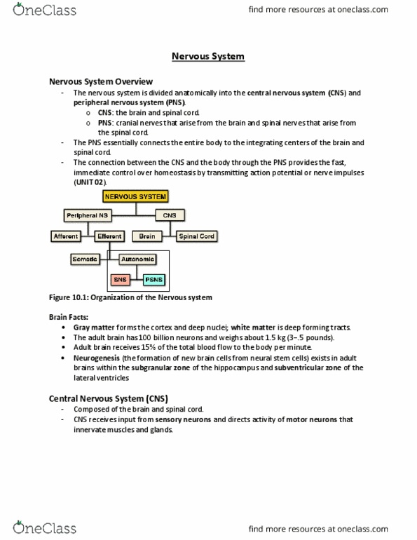
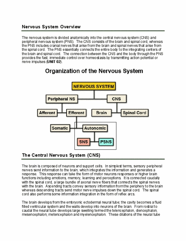
• The nervous system is divided anatomically into:
o CNS (Central Nervous System)
▪ Consist of the brain and spinal nerves that arise from the spinal cord
o PNS (Peripheral Nervous System)
▪ Connects the entire body to the integrating centers of the brain and spinal
cord
▪ Includes:
• Afferent Neurons (sensory)
• Efferent Neurons (motor)
o Somatic
Autonomic (SNS or PSNS)
• The connection between the CNS and the body through the PNS provides fast,
immediate control over homeostasis by transmitting action potential or nerve impulses
The CNS:
• The brain is composed of neurons and support cells
• The sensory peripheral nerves send information to the brain which integrates the
information to generate a response
o Responses can take form of motor neuron responses or higher brain functions
(emotion, memory, learning and perceptions)
• The brain is connected caudally to the spinal cord (a large bundle of axon fibers connect)
o Ascending tracts covey sensory information from the periphery to the brain
o Descending tracts send motor nerve impulses down the spinal cord
• Spinal cord performs some information integration in the form of reflex arcs
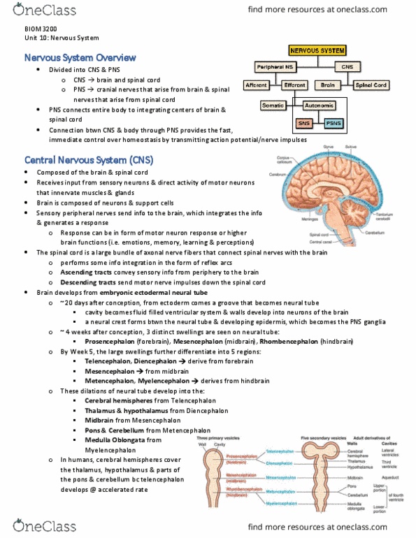
• Development:
o The brain develops from the embryonic ectodermal neural tube
▪ Cavity becomes a fluid filled ventricular system; walls develop into the
neurons of the brain
o From rostral to caudal, the neural tube develops large swelling including:
▪ Telencephalon cerebral hemispheres
▪ Diencephalon thalamus and hypothalamus
▪ Mescencephalon midbrain
▪ Metencephalon pons and cerebellum
▪ Myelencephalon medulla oblongata
find more resources at oneclass.com
find more resources at oneclass.com

2
o In humans, the telencephalon develops at an accelerated rate and results in the
cerebral hemisphere covering the thalamus, hypothalamus and parts of the
pons/cerebellum
Lobe
Functions
Frontal
• Voluntary motor control of skeletal muscles
• Higher intellectual processing
• Verbal communication
Parietal
• Somatesthetic interpretation
• Understanding speech and formulating words to express thoughts
• Interpretation of textures and shapes
Temporal
• Interpretation of auditory sensations
• Storage of auditory and visual experiences
Occipital
• Integration of movements in focusing the eye
• Correlation of visual images with previous experiences and other
sensory stimuli
• Conscious perception of vision
Insula
• Memory
• Sensory and visceral integration
The Ventricles:
• Cavities of the neural tube remain as cavities during development of the mammalian
brain
o As a result, an adult brain contains fluid filled chambers ventricles
• Ventricles are filled with cerebral spinal fluid (CSF)
o This fluid is produced by specialized tissue found in each ventricle choroid
plexus
o CSF provides nourishment and protection as a shock absorber
• The large, paired ventricles are found deep in the cerebral hemispheres and join caudally
on the midline to the third ventricle (at the level of the diencephalon)
• At the level of the midbrain, the ventricle narrows into the aqueduct
o The aqueduct communicates with the fourth ventricle at the level of the pons,
cerebellum and medulla
• The ventricle system continues into the spinal cord as the central canal
• The CSF escapes the ventricles through small openings called foramen into the
subarachnoid space
find more resources at oneclass.com
find more resources at oneclass.com

3
The Meninges:
• The brain and spinal cord are encased in three connective tissue layers = meninges
o Outermost: dura mater (tough connective tissue)
o Middle: arachnoid mater (delicate membrane)
o Innermost: pia mater (delicate membrane that is associated with the surface of
the brain)
• The space between the arachnoid mater and the pia mater is the subarachnoid space
that is filled with CSF
o The purpose of the subarachnoid space is to provide a buoyant layer that protects
the brain from mechanical damage
• Cerebrospinal fluid produced by the choroid plexuses of the ventricles escaped through
small windows or foramen located in the lateral ventricles and the fourth ventricle and
completely surrounds the brain and spinal cord
o CSF in subarachnoid space drains through specialized areas of the meninges
(arachnoid villi) into venous circulation
o Therefore, the brain and spinal cord are continuously bathed and floated in a
protective layer of CSF
Cerebrum:
• Higher brain functions arise in the cerebrum
o It is a large mushroom shaped wrinkle structure found cranially and partially
covering the rest of the brain
o Characterized by grooves called sulci, which form elevated folds =gyri
• The surface of the cerebrum is grey and consists of cell bodies
o Deep into the cerebrum is white matter that consists of myelinated axons that
connect the grey matter with other areas of the brain
o Cell bodies found in white matter often collect in groups and form distinct grey
areas termed nuclei
• Can be divided anatomically into the left and right hemispheres by a deep groove called
the longitudinal fissure
o Each area can then be divided and are labeled to the corresponding skull bones
that overlay each area
• The central sulcus divides the cerebrum coronally into anterior and posterior parts
o It also marks the division of the frontal and parietal lobes
▪ Lateral sulcus is a large groove on each side of the brain and delineated
the temporal lobe from the frontal and parietal lobes
▪ Posterior part = occipital lobe
• Each area corresponds to a specific function
o Precentral gyrus (part of the frontal lobe adjacent to the central sulcus)
find more resources at oneclass.com
find more resources at oneclass.com
Document Summary
In the textbook: figures 8. 5, 8. 6 and 9. 11; tables 8. 1, 9. 1 and 9. 6. Includes: afferent neurons (sensory, efferent neurons (motor, somatic. Autonomic (sns or psns: the connection between the cns and the body through the pns provides fast, immediate control over homeostasis by transmitting action potential or nerve impulses. Functions: voluntary motor control of skeletal muscles, higher intellectual processing, verbal communication, somatesthetic interpretation, understanding speech and formulating words to express thoughts, storage of auditory and visual experiences, correlation of visual images with previous experiences and other. Integration of movements in focusing the eye sensory stimuli: conscious perception of vision, memory, sensory and visceral integration. Limbic system: consists of various structures located in both the telencephalon and diecephalon, including, hippocampus implicated in memory, amygdala, septal nuclei, hypothalamus. In all mammals, the system is important in processing olfactory sensory information. The mesencephalon: (or midbrain) forms the upper part of the brain stem which connects the pons and cerebellum with the diencephalon.