BIOC34H3 Lecture Notes - Lecture 3: Atrial Flutter, Ventricular Fibrillation, Purkinje Fibers

Document Summary
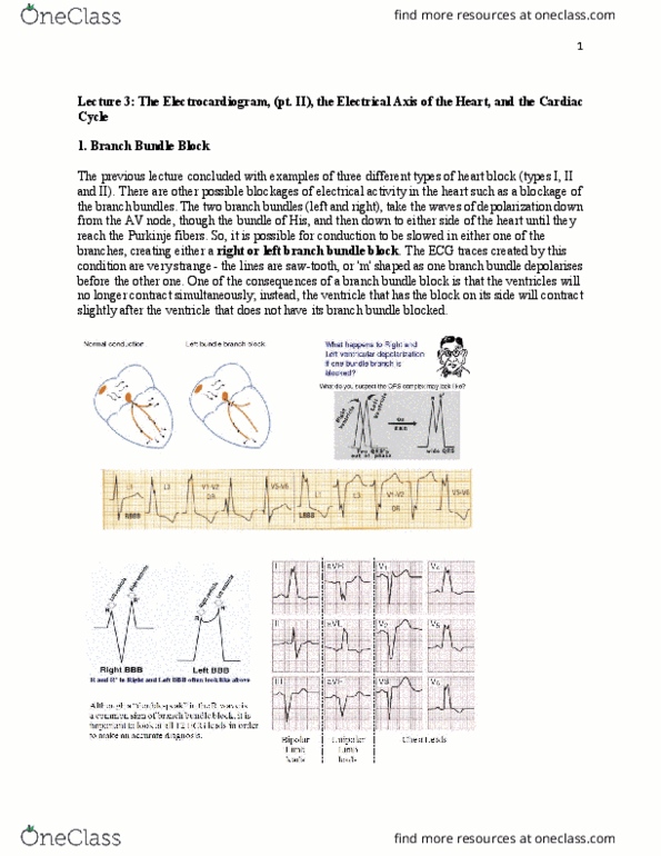
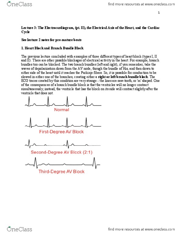
Left and right bundle take electrical activity (waves of depol) from the av node, through the bundle of his, down to the left and right sides of the heart, into the purkinje fibres. Blocking the branch bundles will slow down heart activity/contraction. Ecg trace: saw-tooth or m shaped lines. Ventricles don"t contract completely (only starts to, but then does not finish) Accurate diagnosis look at all 12 ecg patterns: 3 bipolar limp leads (i, ii, iii, 3 unipolar limb leads (avr, avl, avf, 6 chest leads(v1-v6) Usually an m peak at qrs complex. Ecg traces can make the isoelectric level between. Atrial: if the qrs complex remains intact ventricles contract properly. Atrial flutter: rapid rate of electrical excitation enhanced number of p- waves. Atria contracts and depolarizes at diff times. Atrial contraction doesn"t matter as much when the hearts at rest.