PSL301H1 Lecture Notes - Lecture 7: Purkinje Fibers, Intercalated Disc, Sinoatrial Node


41
PSL301H1 Full Course Notes
Verified Note
41 documents
Document Summary
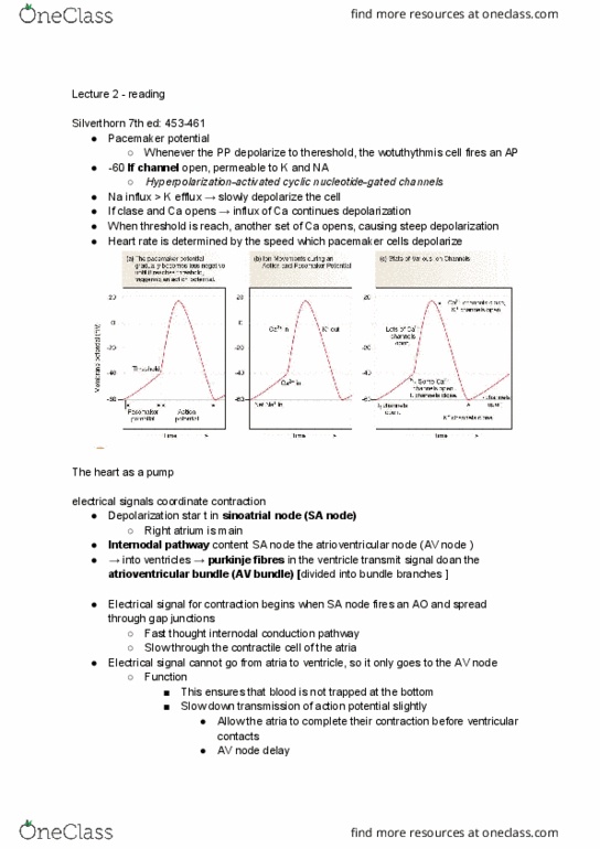
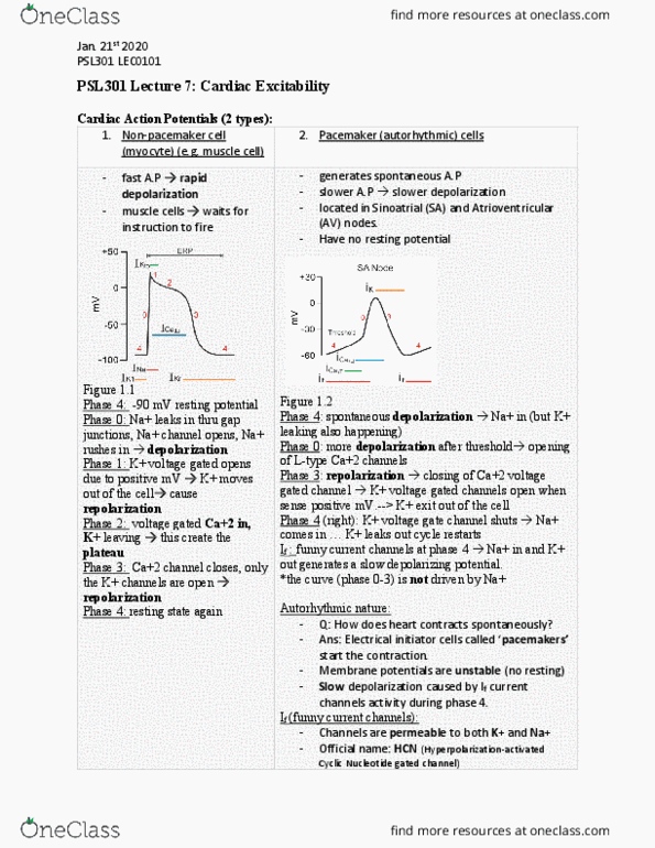
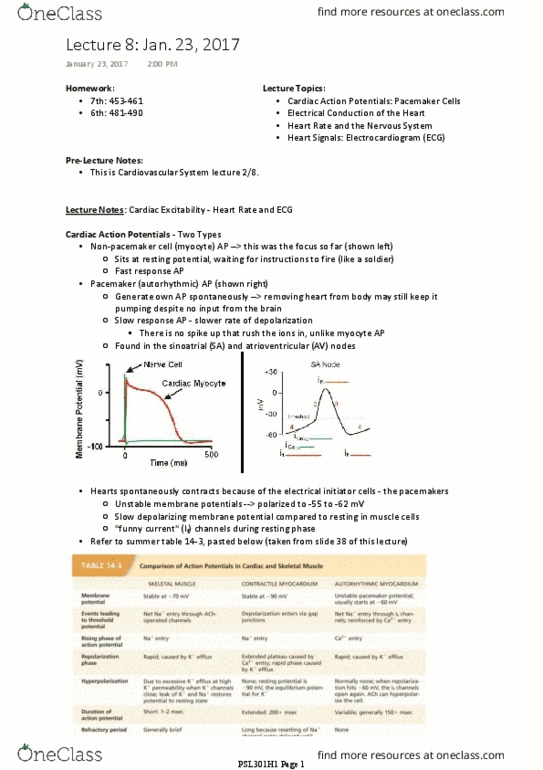
Myocyte: type 1 cardiac ap, non-pacemaker cell: fast response aps, rapid depolarization, muscle cell soldiers" need instructions to fire, ca causes plateau of ap. Internodal pathway: branched fibres connecting sa and av node. Av node: routes direction of electrical signals: delays transmissions of aps, ensures atria complete contraction before ventricles, located at floor of right atrium, can act as pacemakers when sa slow, 50 bpm. Purkinje fibres: specialized conducting cells in ventricles: transmit electrical signals very rapidly, can act as pacemakers when sa slow, 25-40 bpm. Av bundle/bundle of his: fibres of ventricular septum: receives signal from purkinje fibres, divide into l/r bundle branches, travel to apex of heart, divide into smaller purkinje fibres that spread outwards among contractile cells. Electrocardiogram: extracellular recording of ap sums in many heart muscle cells. Pr segment: conduction through av node/bundle, atria contract. P not triggering qrs = 2nd degree block at av node.