Anatomy and Cell Biology 3309 Lecture Notes - Lecture 33: Retinal Pigment Epithelium, The Eye 2, Optic Nerve

2 May 2018
School
Department
Professor

The Eye 2
Objectives
- Understand the organization of the retina
- Compare and contrast rod and cone photoreceptor cells
- Understand the overview of the retinal circuitry
- Understand what fovea, macula and optic disk
The Eye
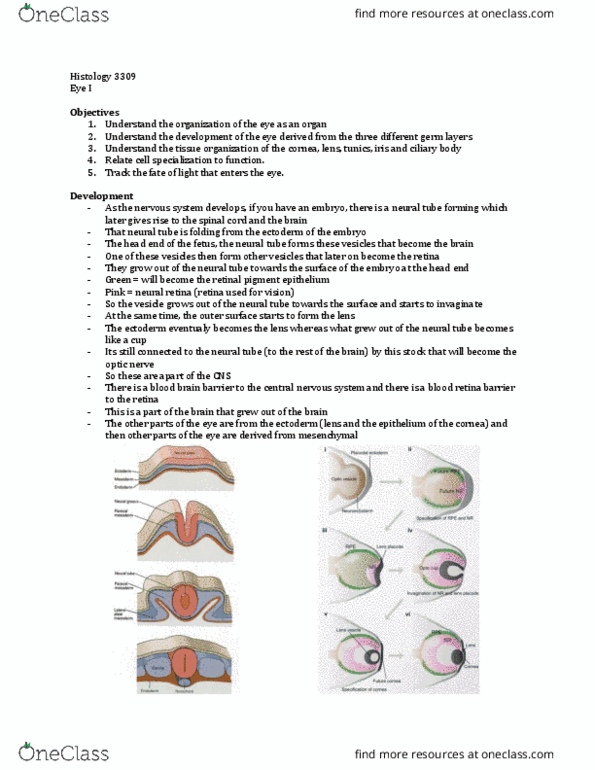
- Optic nerve is continuous with the retina
o Most inner tunic of the 3 tunics of the eye
- Retina comprises of different layers:
o Neural retina (real retina) tissues that contains the photoreceptors that we use to see
o Retinal pigment epithelium next outer layer to the neural retina
o Continuous with the two epithelium layers that cover the ciliary body and the iris
▪ Note: the outer layer of the two epithelium is pigmented and is continuous with the
retinal pigment epithelium
find more resources at oneclass.com
find more resources at oneclass.com

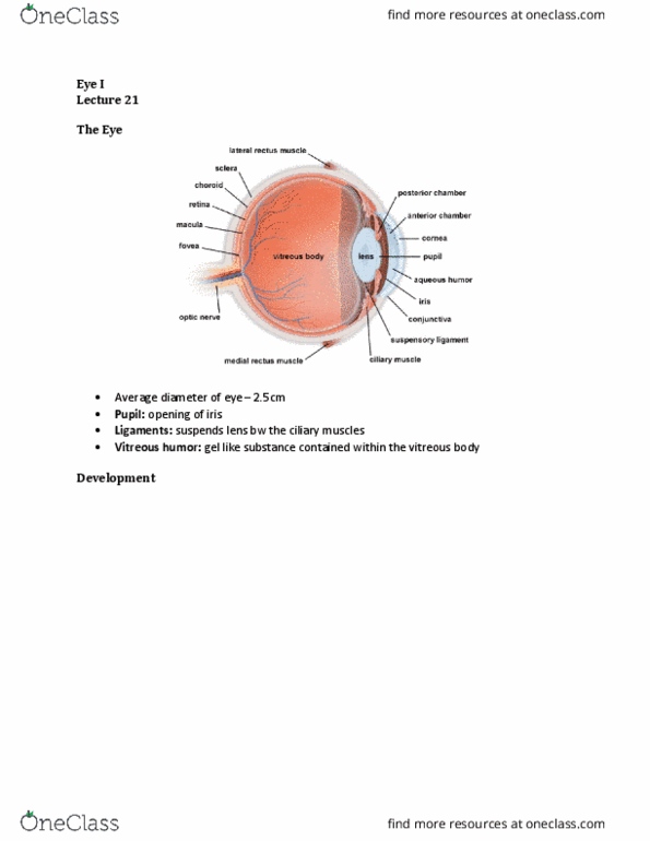
- RETINA!
- Many layers
o Easy to study different type of neurons and how they are connected (nicely organized)
- Pink layer at bottom = retinal pigment epithelium (outer layer) one layer of epithelial cell
o White layer underneath it is the next tunic layer = choroid
▪ Contains all of the blood vessels
o Sclera is after the choroid
- Different layers of cell bodies = nuclear layers
- Layers with no cell bodies = plexiform layers (where the neurons make contact)
o Neuropil =Dendrites, synapses, etc.
- The white top is the centre of the eye
o Where the light comes from
o The photoreceptors are first purple layer from bottom (cell bodies of the photoreceptors)
▪ Outer segment of the photo receptors = pink layer under it. This is where
phototransduction takes place. Light travels through the entire retina in order to hit
the outer segment to induce a neuronal signal
- Red dots in the image = blood vessels
o There are blood vessels in the INNER portion of the retina but NOT the outer portion. No
vascularization in the outer portion of the retina, they have to be provided nutrients from
the choroid and pigment epithelial cells to the outer segments of the receptors
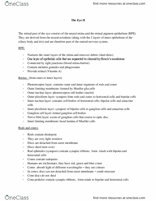
The Retina
- Outer nuclear layer = bodies of the photoreceptors
- Outer plexiform layer
o Synapses of photoreceptors to next cells are here
- Inner nuclear layer – different types of cells
o Horizontal, bipolar, amacrine cells
- Inner plexiform layer
o No cell bodies, only contacts
find more resources at oneclass.com
find more resources at oneclass.com

- Ganglion cell layer:
o Cell bodies of the retinal ganglion cells lie here
▪ Ganglion cells (ONLY CELLS THAT FIRE ACTION POTENTIALS IN RETINA) fire AP,
and their axons travel through the nerve fiber layer all the way to the optic disk, and
merge to the optic nerve and project into the brain
o Axons of the ganglion cells project into the brain
The Retinal Pigment Epithelium (RPE)
- Most outer layer = retinal pigment epithelial cells
o Border to the choroid
- They have a lot of different roles
o 1. Pigmented – contain melanocytes, which means they absorb light. All the light that has not
been absorbed at the photoreceptor cells has to be absorbed somewhere or else it would
enter the brain or the skull
▪ Choroid absorbs some as well
▪ But the RPE contains melanocytes for light absorption.
▪ In some animals, RPE reflects light not absorbs it (so they can see at night)
• NOCTURNAL ANIMALS
o 2. Epithelial transport
▪ Photoreceptor cells are neurons, and have to maintain homeostasis in terms of ion
concentration.
o 3. Glial cell function
▪ Provide nutrients
find more resources at oneclass.com
find more resources at oneclass.com
Document Summary
Compare and contrast rod and cone photoreceptor cells. Understand the overview of the retinal circuitry. Understand what fovea, macula and optic disk. Optic nerve is continuous with the retina: most inner tunic of the 3 tunics of the eye. Many layers: easy to study different type of neurons and how they are connected (nicely organized) Pink layer at bottom = retinal pigment epithelium (outer layer) one layer of epithelial cell: white layer underneath it is the next tunic layer = choroid, contains all of the blood vessels, sclera is after the choroid. Different layers of cell bodies = nuclear layers. Layers with no cell bodies = plexiform layers (where the neurons make contact: neuropil =dendrites, synapses, etc. The white top is the centre of the eye: where the light comes from, the photoreceptors are first purple layer from bottom (cell bodies of the photoreceptors, outer segment of the photo receptors = pink layer under it.