Anatomy and Cell Biology 3309 Lecture 45: Urinary System 1

2 May 2018
School
Department
Professor

Histology Lecture 8 – Semester 2
The Urinary System 1
QUESTIONS
Which of the following substances produced by epithelial cells of the gastric mucosa aids in the resorption of
vitamin B12?
a. Gastrin
b. HCl
c. Intrinsic Factor
- Parietal cells secrete them (Hydrochloric acid secreting cells too!)
d. Pepsinogen
e. Mucus
Learning Objectives
- Describe the anatomical organization of the kidney
- List the components of a uriniferous tubule
- Prepare a labeled diagram of a renal corpuscle
- List the components of the urinary filtration barrier
- Name the factors that contribute to ultrafiltration
- Describe the role of mesangial cells in the renal corpuscle.
- Function of urinary system:
o To clear the blood of the waste products of metabolism
o To regulate the concentrations of many constituents of the body fluids
- Urinary system consists of 2 kidneys and excretory passages to store and conduct excreted materials
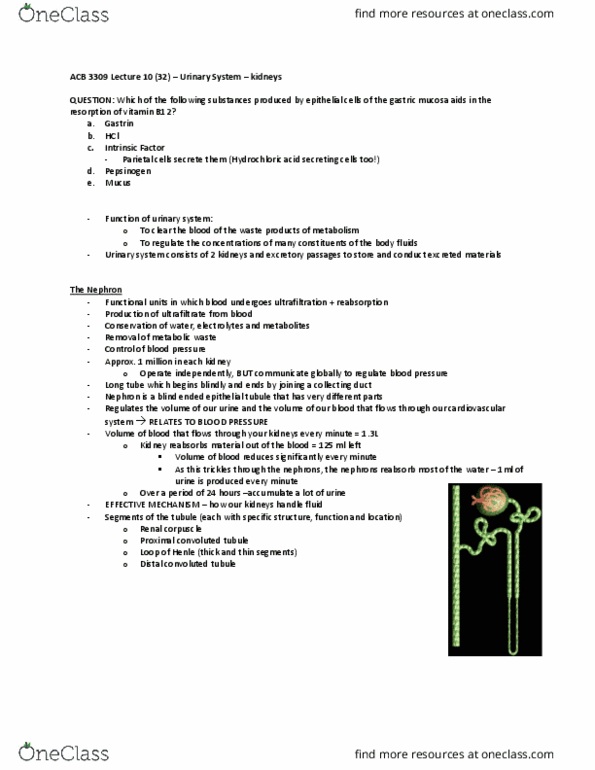
The Nephron
- Functional units in which blood undergoes ultrafiltration + reabsorption
- Production of ultrafiltrate from blood
- Conservation of water, electrolytes and metabolites
- Removal of metabolic waste
- Control of blood pressure
- Approx. 1 million in each kidney
o Operate independently, BUT communicate globally to regulate blood pressure
- Long tube which begins blindly and ends by joining a collecting duct
- Nephron is a blind ended epithelial tubule that has very different parts
o Designed to interact with the blood stream to filter blood and evaluate what is good and
what is not good
o Gets rid of the metabolites (waste products) and recover the good things we want to keep
o Regulates the volume of our urine and the volume of our blood that flows through our
cardiovascular system RELATES TO BLOOD PRESSURE
- Volume of blood that flows through your kidneys every minute = 1300ml
o Kidney resorb material out of the blood = 125 ml left
find more resources at oneclass.com
find more resources at oneclass.com

▪ Volume of blood reduces significantly every minute
▪ As this trickles through the nephrons, the nephrons reabsorb most of the water –
1ml of urine is produced every minute
o Over a period of 24 hours –accumulate a lot of urine
- EFFECTIVE MECHANISM – how our kidneys handle fluid
- Segments of the tubule (each with specific structure, function and location)
o Renal corpuscle
o Proximal convoluted tubule
o Loop of Henle (thick and thin segments)
o Distal convoluted tubule
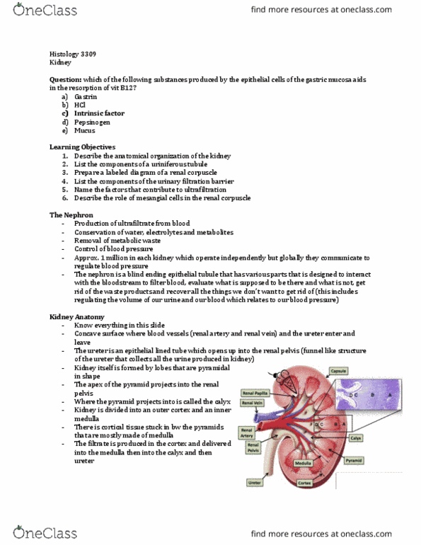
Kidney Anatomy
- KNOW THE PARTS OF THE KIDNEY
- Bean shaped organ
- Lie in retroperitoneal space on the posterior aspect of the abdominal cavity
- Surrounded by capsule, divided into cortex and medulla
- Concave surface where the plumbing goes in and out (blood vessels)
o Renal artery + renal vein enters and leave
o Epithelial lined tube = ureter
▪ Opens up into the renal pelvis (funnel like structure of the ureter that collects all of
the urine that is produced in the kidney)
- Kidney is formed by lobes
o The lobes are organized in pyramidal form (it is a pyramid)
o Pyramid has its apex projecting into the renal pelvis
o Name of the funnel like structure the pyramid projects into = calyx
▪ There are minor and major calyxes
▪ Space where the urine is being delivered into the ureter
- Most kidneys have MANY lobes (humans)
o Some animals (cat) have only ONE lobe
- IMAGE = CAT KIDNEY
o D = pyramid opens up into the calyx
- Kidney is divided into an outer cortex and and inner medulla
o Pyramids structurally are mostly medulla tissue
o Can distinguish these two different parts in low magnification with their staining
▪ THEY LOOK DIFFERENT
▪ Different things are located in the cortex compared to the medulla
- Each pyramid can be likened to an ice cream cone
o Medulla = cone itself
o Cortex = ice cream
o Hot day, the ice cream runs down the cone – HAPPENS IN THE KIDNEY
▪ Cortex dips down in between sections of the medulla (renal columns)
▪ Cortical tissue stuck in between the pyramids (mostly made up of medulla)
- Filtrate is produced in the cortex and delivered through the medulla into the calyx and the ureter
- Each lobe is like a gland, has lobules
o KIDNEY HAS LOBULES!
find more resources at oneclass.com
find more resources at oneclass.com

- Surface features:
o Hilus = concavity on the medial border
o Ureter = large excretory duct which passes from the hilus to the bladder
o Pelvis = the upper expanded end of the ureter which fills the hilus
o Papilla = conical protrusion of renal substance enveloped by a minor calyx
o Collecting duct = 10-25 ducts which perforate each papilla
- Hemisected view:
o Medullary pyramids = 8-12 pyramids in the medulla with tips projecting to the calyx
o Renal columns = cortical tissue extending into the medulla between pyramids
o Medullary rays = medullary material extends into the cortex as fine radial rays
o Lobe = a pyramid with its associated overlying cortex
o Lobule = all nephrons draining into a collecting duct
▪ Has a medullary ray at the centre
▪ Interlobular arteries lie between the lobules
-
Uniferous tubule
- One uniferous tubule includes a nephron and the collecting duct into which it empties
find more resources at oneclass.com
find more resources at oneclass.com
Document Summary
Which of the following substances produced by epithelial cells of the gastric mucosa aids in the resorption of vitamin b12: gastrin, hcl. Parietal cells secrete them (hydrochloric acid secreting cells too!: pepsinogen, mucus. Describe the anatomical organization of the kidney. Prepare a labeled diagram of a renal corpuscle. List the components of the urinary filtration barrier. Name the factors that contribute to ultrafiltration. Describe the role of mesangial cells in the renal corpuscle. Function of urinary system: to clear the blood of the waste products of metabolism, to regulate the concentrations of many constituents of the body fluids. Urinary system consists of 2 kidneys and excretory passages to store and conduct excreted materials. Functional units in which blood undergoes ultrafiltration + reabsorption. 1 million in each kidney: operate independently, but communicate globally to regulate blood pressure. Long tube which begins blindly and ends by joining a collecting duct.