Kinesiology 2230A/B Lecture Notes - Lecture 7: Alpha Motor Neuron, Biceps Femoris Muscle, Intrafusal Muscle Fiber

28 Jun 2017
School
Department
Course
Professor

Document Summary
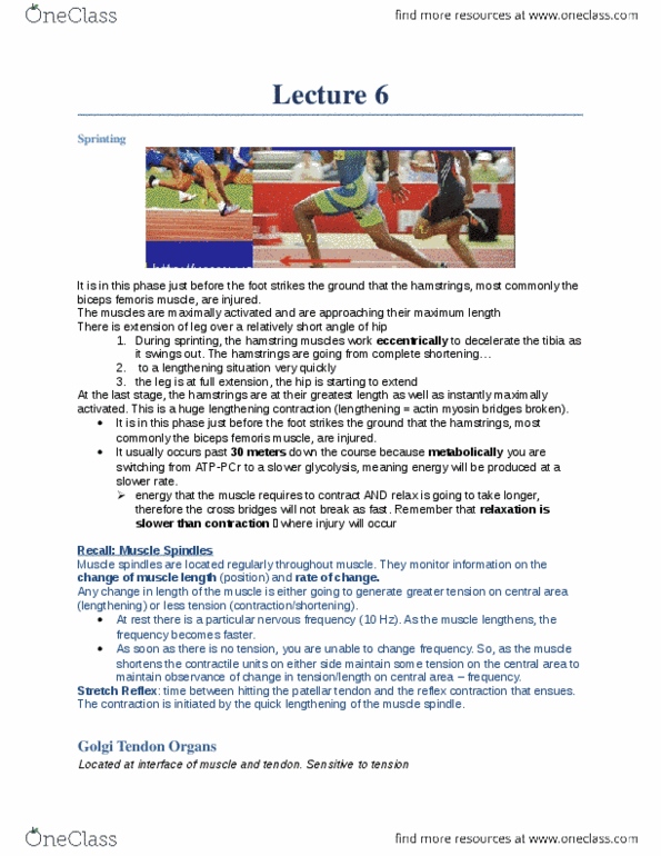
Muscle spindles: contractile areas on either side of the central area (collagen connective tissue) are intrafusal fibers (all the fibers outside the muscle spindle) associated with the whole muscle. When the muscle is shortened, the opposite happens. As a result, we can tell where the limb (or muscle at least) is in space and by noting the rate of change in discharge frequency, we can tell how fast position is changing. Indicate both absolute amount and rate of change: spindles are activated by gamma motor neurons and can assist in rapid movement. Located in that interface between the contractile proteins and the tendons themselves: golgi tendon organs are associated with monitoring change in tension, how much tension is being placed on a muscle; what is the load on the muscle. If you are disrupting membrane structure in the muscle, you will have leakage of the myoglobin and ck in the bloodstream.