Kinesiology 2230A/B Lecture Notes - Lecture 26: Neuromuscular Junction, Endoplasmic Reticulum, T-Tubule

19 Oct 2018
School
Department
Course
Professor
Document Summary
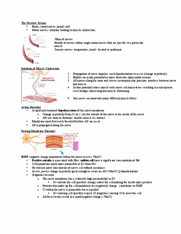
Most sc injuries are shearing of the vertebrae themselves cervical: Can see sc injury very low in the sacrum. If you ever injure the tail bone/coccyx it takes a while to recover. As you move up sc, lose more and more function downstream diagram: Nodes of ranvier are areas that can depolarized. Speed of ap moved more quickly though myelinated axons. Ap moves through axon, ends up at neuromuscular junction. Have calcium voltage gated channels releasing ca into terminal. Have something containing acetyl choline which is release from presence of ca into synaptic cleft. Binds to receptors on the na channels. Once na influx occurs here then propagation of ap is similar than the fibre itself. Have voltage gated sodium channels along the sarcolemma/cell membrane picture: T tubules are perpendicular to muscle fibres/contractile filaments. Ap moves along t tubules which surround all muscle fibres.