BIOL-242 Lecture Notes - Lecture 31: Alveolar Cells, Thoracic Wall, Macrophage

26 May 2020
School
Department
Course
Professor
Document Summary
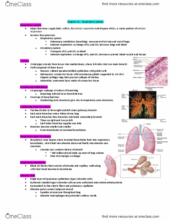
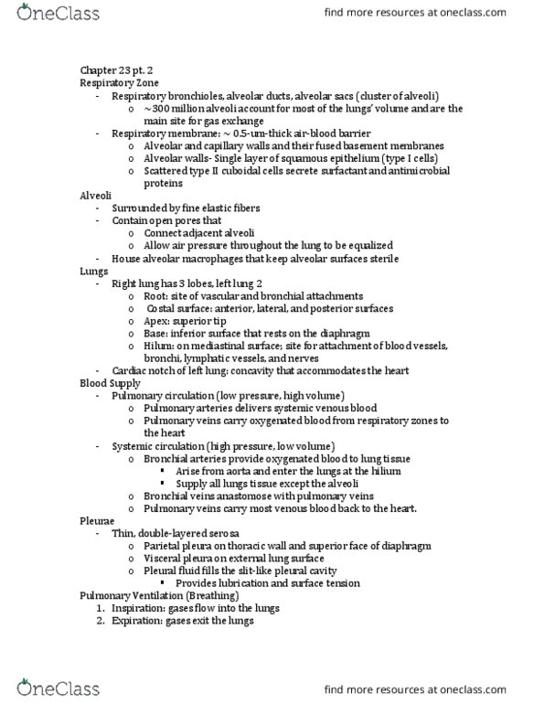
Chapter 22 part 3 | alveoli, lungs, pleurae, mechanics of breathing. Alveolar and capillary walls and their fused basement membranes. ~0. 5- m-thick; gas exchange across membrane by simple diffusion. Single layer of squamous epithelium (type i alveolar cells) Scattered cuboidal type ii alveolar cells secrete surfactant and antimicrobial proteins. Surrounded by fine elastic fibers and pulmonary capillaries. 2 million dead macrophages/hour carried by cilia throat swallowed. Root-site of vascular and bronchial attachment to mediastinum. Costal surface // anterior, lateral, and posterior surfaces. Apex // superior tip; deep to clavicle. Base // inferior surface; rests on diaphragm. Hilum // on mediastinal surface; site for entry/exit of blood vessels, bronchi, lymphatic vessels, and nerves. Separated into superior and inferior lobes by oblique fissure. Superior, middle, inferior lobes separated by oblique and horizontal fissures. Bronchopulmonary segments (10 right, 8 10 left) separated by connective tissue septa. Lobules // smallest subdivisions visible to naked eye; served by bronchioles and their branches.