PSYC 3300 Lecture Notes - Lecture 19: Visual Acuity, Somatotopic Arrangement, Visual Cortex

Document Summary
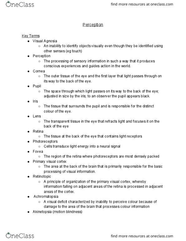
Iris: colored part of the eye: circular muscle controls pupil dilation/constriction, pupil: opening of the eye where light is allowed to enter, cornea: transparent outer layer, refracts light, lens: flexible tissue, inverts image, muscular control of focus. Retinotopic organization: visual cortex contains visual field maps, nearby cells transmit information all the way to the visual cortex, auditory system has tonotopic organization, sensory system has somatotopic organization. The blindspot: the optic nerve exits the eye at the optic disk, creates a blindspot because there are no photoreceptors. In the fovea, there is a 1:1 cone to ganglion cell ratio. Receptive fields: the number of rods increases towards periphery. In the periphery, there is a 1 gc:10prs or 100prs ratio. In the absence of light the na+ and ca+ channels are open. Photopigments and neural signals: receptors are filled with light sensitive chemicals: photopigments, light breaks down the molecules, chemical reaction leads to a neural response, transduction of light into neural energy.