BIO 263 Lecture Notes - Lecture 11: Thoracic Inlet, Chordae Tendineae, Tricuspid Valve


Thorax, Heart, Lungs
Thorax
The border of the thorax is the ribs, the diaphragm is the division between the thorax and
abdomen
- Two holes for esophagus and major blood vessels
Thorax skeletal framework:
- Vertebrae (12)
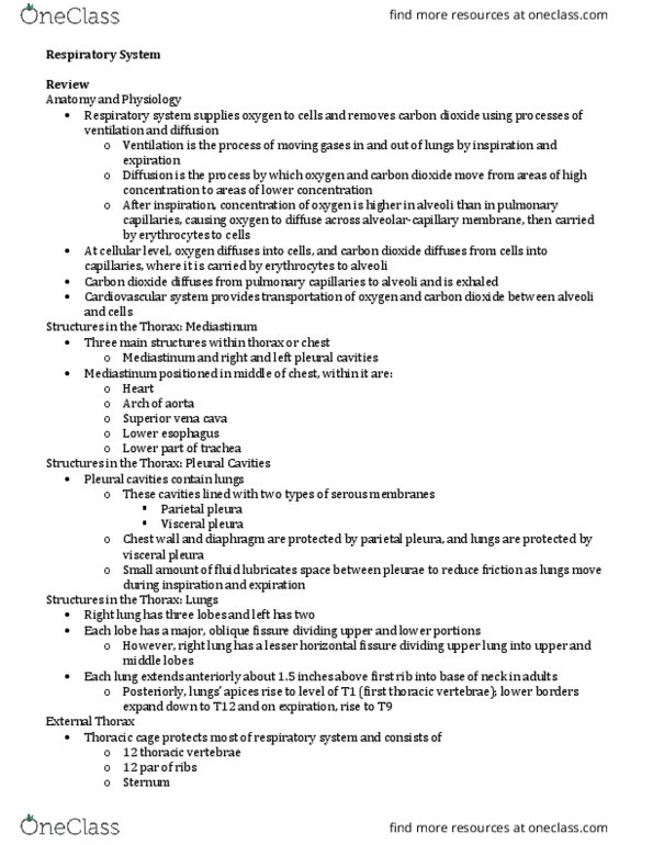
- Ribs (12 pairs), for breathing, but also protect
- Sternum
- Xiphoid process
Intercostal Spaces
- Intercostal Muscles
- Diaphragm
Organs
- Heart
- Lungs (2)
Mediastinum
- Broad central partition that separates the two pleural cavities.
- Borders: Sternum to the vertebrae, superior thoracic aperture to the diaphragm
- Divided into superior, anterior, middle, posterior mediastinum.
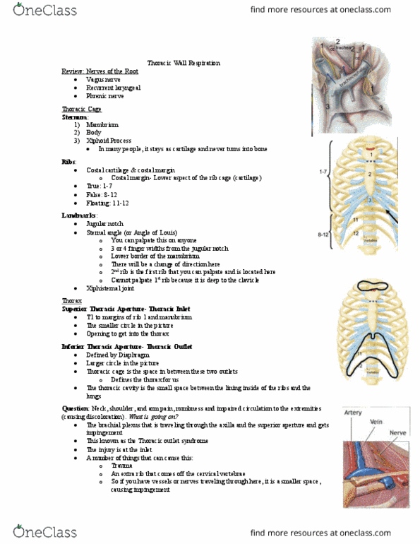
- Superior = major blood vessels to and from heart, and trachea
- Middle = heart
- Posterior = aorta and esophagus
find more resources at oneclass.com
find more resources at oneclass.com

Pericardium
- Fibrous sac that covers the heart and the roots to the great vessels.
Serous pericardium
- Parietal
- Visceral
Pericardial cavity- space between the two layers of the serous membrane
Wedge Analogy:
- Apex is the lowest part, pointing down and left
- Base is flat, backside of heart (blood vessels
interface with heart at the base)
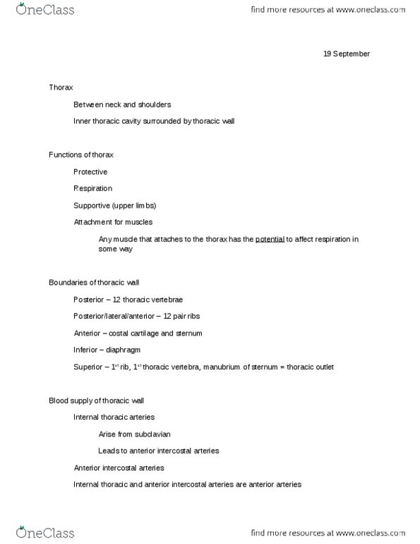
Arteries: carry blood away from the heart
Vein: carries blood to the heart
Valves: keep blood flowing in one direction in the heart
Right Ventricle
- Conus arteriosus: outflow tract (heart → lungs → and back to heart)
- Trabeculae carneae: irregular muscle structures
- Chordae tendineae: connect to free edges of tricuspid valve
find more resources at oneclass.com
find more resources at oneclass.com

Right Atrium
Left Atrium
- Most of base or posterior part of heart
- Openings for pulmonary veins
- Left auricle
- L atrioventricular orifice (bicuspid valve)
Left Ventricle
find more resources at oneclass.com
find more resources at oneclass.com
Document Summary
The border of the thorax is the ribs, the diaphragm is the division between the thorax and abdomen. Two holes for esophagus and major blood vessels. Ribs (12 pairs), for breathing, but also protect. Broad central partition that separates the two pleural cavities. Borders: sternum to the vertebrae, superior thoracic aperture to the diaphragm. Divided into superior, anterior, middle, posterior mediastinum. Superior = major blood vessels to and from heart, and trachea. Fibrous sac that covers the heart and the roots to the great vessels. Pericardial cavity- space between the two layers of the serous membrane. Apex is the lowest part, pointing down and left. Base is flat, backside of heart (blood vessels interface with heart at the base) Valves: keep blood flowing in one direction in the heart. Conus arteriosus: outflow tract (heart lungs and back to heart) Chordae tendineae: connect to free edges of tricuspid valve. Most of base or posterior part of heart.