NURS 301 Lecture Notes - Lecture 4: Somatic Nervous System, Peripheral Nervous System, Autonomic Nervous System


The Nervous System
Sensory input --> Integration --> Motor output
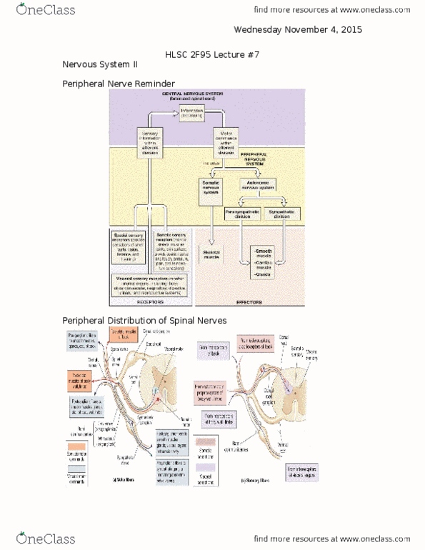
Organization of Nervous System
Central nervous system (afferent)
Brain and spinal cord
○
Where information is integrated
○
Sensory input
Inside and outside our body
§
○
-
Peripheral nervous system (efferent)
Somatic nervous system
Skeletal muscles
§
○
Autonomic nervous system
Work together to create homeostasis
§
Using a lot of energy
§
Parasympathetic
Conservation of energy
□
§
Sympathetic
Smooth muscle, cardiac muscle, glands
□
§
○
-
Receptors (peripheral nervous system)
Special sensory receptors
Provides sensations of smell, taste, vision, balance, or hearing
§
○
Somatic sensory receptors
Monitor skeletal muscles, joints, skin surface, provide position sense and touch, pressure, pain, or temperature sensations
§
○
Visceral sensory receptors
Monitor internal organism including cardiovascular, respiratory, digestive, urinary, reproductive
§
○
-
Sensory neurons afferent
Brings sensory in
○
-
Efferent brings sensory information out
-
"A before E"
-
Histology of Nerve Tissue
Two principle cells of the nervous system
Neurons: excitable cells that transmit electrical signals
Structural units of the nervous system
Composed of a body, axon, and dendrites
□
Long-lived, high metabolic rate
□
§
Their plasma membrane functions in:
Electrical signaling
□
Cell-to-cell signaling during development
□
§
○
Neuroglial: supporting cells
○
-
Functional Classes of Neurons
Sensory (afferent) neurons: detect stimuli
-
Interneurons: receive the information and integrate
-
Motor (efferent) neurons: respond
-
Neuron Structure
Body has all the organelles
-
Dendrites direct communication through the soma
-
Axon hillock where the dendrite arises
-
Myelin Sheath
Whitish, fatty (protein and lipid), segmented sheath around most long axons
○
It functions to
Protect the axon
§
Electrically insulate fibers from one another
§
Increase the speed of nerve impulse transmission
§
○
-
Schwann cell wraps around the axon
300 layers in myelin sheath
○
Cell membrane has a lot of lipid
○
-
Neurilemmal superficial
-
Spots along the axon have nodes between the schwann cells
-
Myelination
Other neuroglial cells in central nervous system
-
Oligodendrocytes
-
Unmyelinated Nerve Fibers
Unmyelinated PNS axons are also surrounded by Schwann cells, but the Schwann cells do not coil densely around these axons
○
-
Structural Classes of Neurons
Anaxonic neuron
Looks like starburst
○
Covers the brain
○
Small extensions
○
-
Bipolar neuron
1 side has the dendrites, 1 has axon
○
-
Unipolar neuron
One long extension wit body coming off
○
-
Multipolar neuron (99% of neurons)
2 or more dendrites
○
Single axon coming off
○
-
Neuroglia
Structural and protection: provide a supportive scaffolding for neurons; segregate and insulate neurons; guide young neurons to the
proper connections
-
Adjust the chemical environment around neurons for health and growth
-
Some produce cerebral fluid: salty solution that the CNS is bathed in
-
In peripheral nervous system
Schwann cells: surrounds all axons in PNS, responsible for myelination of peripheral axons; participate in repair process after
injury
○
Satellite cells: surround neuron cell bodies in ganglia; regulate oxygen and carbon dioxide, nutrient and neurotransmitter levels
around neurons in ganglia
○
-
In central nervous system
Oligodendrocytes: myelinate CAN axons; provide structural framework
○
Astrocytes: maintain brain-blood barrier
Structural support
§
Regulate ion, nutrient, dissolved gas, concentrations
§
Most prevalent
§
○
Microglia: remove cells debris, waste, and pathogens
○
Ependymal cells: line ventricles and central canal, assist in producing, circulating, and monitoring CBS fluid
○
-
Regions of the Brain and Spinal Cord
White matter: dense collections of myelinated fibers
-
Gray matter: mostly soma (cell bodies) and unmyelinated fibers
-
Peripheral nervous system
Gray matter - ganglia
○
White matter - nerves
○
-
Central Nervous system
Gray matter - neural cortex, centers, nuclei, higher centers
○
White matter - tracts, columns
○
-
Central canal
-
How do Neurons communicate?
Communication by Action Potential
Resting potential: uneven distribution of ions across a living membrane
○
Stimulus disturbance or change that may lead to an action potential
○
Wave of change in membrane potential:
Depolarization: Na+ positive diffusion into axon
§
Repolarization: K+ positive diffusing out of axon
§
○
Back to resting potential
○
-
Propagation of an Action Potential
More sodium in, and potassium channels out
○
-
Saltatory Propagation
"To be"
○
Action potential moves from node to node
○
Faster to be myelinated
○
-
Signal Conduction
Signal conduction speed depends on two factors
Diameter of fiber
Larger are faster
□
Less resistance will allow it to move faster
□
§
Presence of myelin
Myelinated are faster
□
§
○
Fastest fibers are both large and myelinated
○
-
If neurons are damaged can they be repaired?
YES
-
If cell body remains intact, cut nerve fibers can regenerate
-
Schwann cells secrete nerve growth factors
-
Schwann cells and endoneurium produce regeneration tube to direct regrowth of axon
-
CNS neurons cannot regenerate
-
Damaged neuron in PNS
Step 1: fragmentation of axon and myelin occurs in distal stump
○
Step 2: Schwann cells form cord, grow into cut and unite stumps
Macrophages engulf degenerated axon and myelin
§
○
Step 3: axon sends buds into network of Schwann cells and then starts growing along cord of Schwann cells
○
Step 4: Axon continues to grow and is enfolded by Schwann cells
○
-
How does the message get from one neuron to another in a circuit?
Synapses: meeting point of neuron and other cell
Recall: neuromuscular junction
○
Presynaptic neuron to postsynaptic neuron
○
Axon may connect with the dendrites, soma, or axon of another neuron
○
-
Chemical synapse
Presynaptic neuron releases neurotransmitter to postsynaptic cell
○
Neurotransmitters: messenger molecules
Some are excitatory, some are inhibitory
§
○
Structure at synapse
Synaptic knob of presynaptic cell
Contain synaptic vesicles: packets of neurotransmitters
□
§
Synaptic cleft = space
§
Neurotransmitter receptors on postsynaptic cell
§
○
-
What happens to the neurotransmitter left behind in the synaptic cleft?
Reuptake of the neurotransmitter and recycle it
-
Or degraded
-
Presynaptic Inhibition
Reduces potential
-
Or increased effect on postsynaptic membrane
-
Electrical Synapse
Adjacent cells joined by gap junctions
-
Ions diffuse from cell to cell
-
Quick transmission
-
No integration or decision making
-
Neuronal circuits and pools
From simple to extremely complex
-
Neuronal pools = functional groups of neurons consisting of thousands of neurons including inhibitory and excitatory neurons
-
Patterns of synaptic connections between neurons in pools of neurons called circuits
Diverging
○
Converging
○
Reverberating - pattern
E.g. breathing
§
○
Parallel after discharge
○
-
SOMA = body
Week 4 - 4/16
Monday, April 16, 2018
3:34 PM

The Nervous System
Sensory input --> Integration --> Motor output
Organization of Nervous System
Central nervous system (afferent)
Brain and spinal cord
○
Where information is integrated
○
Sensory input
Inside and outside our body
§
○
-
Peripheral nervous system (efferent)
Somatic nervous system
Skeletal muscles
§
○
Autonomic nervous system
Work together to create homeostasis
§
Using a lot of energy
§
Parasympathetic
Conservation of energy
□
§
Sympathetic
Smooth muscle, cardiac muscle, glands
□
§
○
-
Receptors (peripheral nervous system)
Special sensory receptors
Provides sensations of smell, taste, vision, balance, or hearing
§
○
Somatic sensory receptors
Monitor skeletal muscles, joints, skin surface, provide position sense and touch, pressure, pain, or temperature sensations
§
○
Visceral sensory receptors
Monitor internal organism including cardiovascular, respiratory, digestive, urinary, reproductive
§
○
-
Sensory neurons afferent
Brings sensory in
○
-
Efferent brings sensory information out
-
"A before E"
-
Histology of Nerve Tissue
Two principle cells of the nervous system
Neurons: excitable cells that transmit electrical signals
Structural units of the nervous system
Composed of a body, axon, and dendrites
□
Long-lived, high metabolic rate
□
§
Their plasma membrane functions in:
Electrical signaling
□
Cell-to-cell signaling during development
□
§
○
Neuroglial: supporting cells
○
-
Functional Classes of Neurons
Sensory (afferent) neurons: detect stimuli
-
Interneurons: receive the information and integrate
-
Motor (efferent) neurons: respond
-
Neuron Structure
Body has all the organelles
-
Dendrites direct communication through the soma
-
Axon hillock where the dendrite arises
-
Myelin Sheath
Whitish, fatty (protein and lipid), segmented sheath around most long axons
○
It functions to
Protect the axon
§
Electrically insulate fibers from one another
§
Increase the speed of nerve impulse transmission
§
○
-
Schwann cell wraps around the axon
300 layers in myelin sheath
○
Cell membrane has a lot of lipid
○
-
Neurilemmal superficial
-
Spots along the axon have nodes between the schwann cells
-
Myelination
Other neuroglial cells in central nervous system
-
Oligodendrocytes
-
Unmyelinated Nerve Fibers
Unmyelinated PNS axons are also surrounded by Schwann cells, but the Schwann cells do not coil densely around these axons
○
-
Structural Classes of Neurons
Anaxonic neuron
Looks like starburst
○
Covers the brain
○
Small extensions
○
-
Bipolar neuron
1 side has the dendrites, 1 has axon
○
-
Unipolar neuron
One long extension wit body coming off
○
-
Multipolar neuron (99% of neurons)
2 or more dendrites
○
Single axon coming off
○
-
Neuroglia
Structural and protection: provide a supportive scaffolding for neurons; segregate and insulate neurons; guide young neurons to the
proper connections
-
Adjust the chemical environment around neurons for health and growth
-
Some produce cerebral fluid: salty solution that the CNS is bathed in
-
In peripheral nervous system
Schwann cells: surrounds all axons in PNS, responsible for myelination of peripheral axons; participate in repair process after
injury
○
Satellite cells: surround neuron cell bodies in ganglia; regulate oxygen and carbon dioxide, nutrient and neurotransmitter levels
around neurons in ganglia
○
-
In central nervous system
Oligodendrocytes: myelinate CAN axons; provide structural framework
○
Astrocytes: maintain brain-blood barrier
Structural support
§
Regulate ion, nutrient, dissolved gas, concentrations
§
Most prevalent
§
○
Microglia: remove cells debris, waste, and pathogens
○
Ependymal cells: line ventricles and central canal, assist in producing, circulating, and monitoring CBS fluid
○
-
Regions of the Brain and Spinal Cord
White matter: dense collections of myelinated fibers
-
Gray matter: mostly soma (cell bodies) and unmyelinated fibers
-
Peripheral nervous system
Gray matter - ganglia
○
White matter - nerves
○
-
Central Nervous system
Gray matter - neural cortex, centers, nuclei, higher centers
○
White matter - tracts, columns
○
-
Central canal
-
How do Neurons communicate?
Communication by Action Potential
Resting potential: uneven distribution of ions across a living membrane
○
Stimulus disturbance or change that may lead to an action potential
○
Wave of change in membrane potential:
Depolarization: Na+ positive diffusion into axon
§
Repolarization: K+ positive diffusing out of axon
§
○
Back to resting potential
○
-
Propagation of an Action Potential
More sodium in, and potassium channels out
○
-
Saltatory Propagation
"To be"
○
Action potential moves from node to node
○
Faster to be myelinated
○
-
Signal Conduction
Signal conduction speed depends on two factors
Diameter of fiber
Larger are faster
□
Less resistance will allow it to move faster
□
§
Presence of myelin
Myelinated are faster
□
§
○
Fastest fibers are both large and myelinated
○
-
If neurons are damaged can they be repaired?
YES
-
If cell body remains intact, cut nerve fibers can regenerate
-
Schwann cells secrete nerve growth factors
-
Schwann cells and endoneurium produce regeneration tube to direct regrowth of axon
-
CNS neurons cannot regenerate
-
Damaged neuron in PNS
Step 1: fragmentation of axon and myelin occurs in distal stump
○
Step 2: Schwann cells form cord, grow into cut and unite stumps
Macrophages engulf degenerated axon and myelin
§
○
Step 3: axon sends buds into network of Schwann cells and then starts growing along cord of Schwann cells
○
Step 4: Axon continues to grow and is enfolded by Schwann cells
○
-
How does the message get from one neuron to another in a circuit?
Synapses: meeting point of neuron and other cell
Recall: neuromuscular junction
○
Presynaptic neuron to postsynaptic neuron
○
Axon may connect with the dendrites, soma, or axon of another neuron
○
-
Chemical synapse
Presynaptic neuron releases neurotransmitter to postsynaptic cell
○
Neurotransmitters: messenger molecules
Some are excitatory, some are inhibitory
§
○
Structure at synapse
Synaptic knob of presynaptic cell
Contain synaptic vesicles: packets of neurotransmitters
□
§
Synaptic cleft = space
§
Neurotransmitter receptors on postsynaptic cell
§
○
-
What happens to the neurotransmitter left behind in the synaptic cleft?
Reuptake of the neurotransmitter and recycle it
-
Or degraded
-
Presynaptic Inhibition
Reduces potential
-
Or increased effect on postsynaptic membrane
-
Electrical Synapse
Adjacent cells joined by gap junctions
-
Ions diffuse from cell to cell
-
Quick transmission
-
No integration or decision making
-
Neuronal circuits and pools
From simple to extremely complex
-
Neuronal pools = functional groups of neurons consisting of thousands of neurons including inhibitory and excitatory neurons
-
Patterns of synaptic connections between neurons in pools of neurons called circuits
Diverging
○
Converging
○
Reverberating - pattern
E.g. breathing
§
○
Parallel after discharge
○
-
SOMA = body
Week 4 - 4/16
Monday, April 16, 2018 3:34 PM
Document Summary
Provides sensations of smell, taste, vision, balance, or hearing. Monitor skeletal muscles, joints, skin surface, provide position s. , or hearing position sense and touch, pressure, pain, or temperature sensations piratory, digestive, urinary, reproductive. Whitish, fatty (protein and lipid), segmented sheath around most long a. Schwann cell wraps around the axon piratory, digestive, urinary, reproductive ost long axons. Spots along the axon have nodes between the schwann cells. Unmyelinated pns axons are also surrounded by schwann cells, but. 1 side has the dendrites, 1 has axon. Structural and protection: provide a supportive scaffolding for neurons proper connections. Adjust the chemical environment around neurons for health and growth. Some produce cerebral fluid: salty solution that the cns is bathed in. Schwann cells: surrounds all axons in pns, responsible for myelina injury. Satellite cells: surround neuron cell bodies in ganglia; regulate oxyge around neurons in ganglia. Ependymal cells: line ventricles and central canal, assist in produc.