NURS 301 Lecture Notes - Lecture 7: Subclavian Vein, Lymph Node, Internal Jugular Vein


Lymphatic system consists of
Network of lymphatic vessels
1.
Lymph
Basically interstitial fluid
○
"clear" like plasma
○
2.
Lymphoid tissues
3.
Lymphoid organs
4.
Functions of Lymphatic System
Returning fluid and proteins filtered out of the capillaries into
the circulatory system
-
Picking up fat absorbed at the small intestine and lipid-soluble
vitamins transferring them into the circulatory system
Allows absorption of fats
○
Capillaries can't pick up large fats
Lymph picks up large fats and dumbs them into
circulatory system
§
○
-
Serving as a filter to help capture and destroy foreign pathogens
Pathogens: organisms that cause disease, infectious agents
that enter body
○
Lymphatic system needs to filter them out
○
-
Lymphatic Vessels
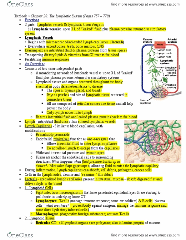
Lymphatic vessels form a one way system in which lymph
flows to the heart
-
There is no pumping of the lymph
-
Series of vessels going from tiny microscopic into larger and
larger vessels
Vessels carry lymph back towards heart and dump
intravenous side of circulatory system
○
-
Cells of Lymphatic System
Lymphocytes, type of white blood cells
White blood cells defend against pathogens
○
Leukocytes, remove pathogens
○
-
Other leukocytes
Macrophages, destroy and engulf foreign invaders
○
-
Cell that secrete connective tissue matrix
Reticulocytes
○
-
Lymphatic capillaries wind around tissue cells and blood
capillaries
Intertwined around tissue cells and blood capillaries
○
Lymph capillaries are blunt ended
Have trap door like structures, cells are overlapping
§
String like collage fibers that hold onto little flaps
§
When fluid builds up, it pushes on the lymph
§
Goes into larger vessels and eventually blood supply
§
○
-
These are wide-spread, except not found in CNS or bone
marrow, teeth
-
Valves in the Lymphatic Vessels
Prevent backflow of lymph
-
Doesn't cool
-
Lymphatic Vessels
Lymphatic collecting vessels with valves are large vessels that
move through the lymph nodes, same three tunics as veins but
thinner-walled and more valves
-
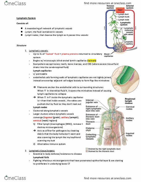
Lymphatic trunks form from the union of largest collecting
vessels. Major trunks are named for the areas of the body they
drain: lumbar, bronchomediastinal, subclavian, jugular,
intestinal
-
Lymphatic ducts the largest of the lymphatic vessel dumps
lymph into the circulatory system
Right lymphatic duct
Dump lymph into between internal jugular and right
subclavian vein
§
Dump into venous side or circulatory system
§
Half inch long
§
Drains from right arm area and thoracic area of right
side and right side of head
§
○
Thoracic duct on left side of body
Draining lymph from rest of the body
§
About 15 inches long
§
○
-
Lymph formation and transport
Excess interstitial fluid (about 3 liters/day); any proteins that
leave plasma cannot return to the blood but can enter lymphatic
capillaries
-
Factors affecting blood flow in veins also apply to lymphatic
vessels
Skeletal contractions, squeezes lymph, cannot cool down
○
-
More activity ---> increase flow into lymphatic system,
balancing the increase rate of fluid loss from the blood
More fluid in capillaries, active tissue
Needs a lot of oxygen for fluid
§
○
-
Can be more than 3 liters a day depending on amount of
activity
-
Edema due to Filarial Infection
Blockage due to parasites, tumors, or removal of lymphatic
tissues results in edema
-
Infection delivered through bite of mosquito
-
Lymph cannot be drained
Puts pressure on nerves, and causes damage to tissue
○
-
Lymphoid Tissues
Lymphoid tissues are distributed throughout the body
-
Diffused lymphocytes or more clustered
Nodules of lymphocytes
○
No outer capsule, wouldn't call it an organ
○
-
Found in mucus membranes or organs
-
MALT = mucosa-associated lymphatic tissue
Mucous membrane association
○
E.g. Peyer's patch
Found in small intestines
§
Involved in monitoring for any foreign invaders
§
○
-
Lymphoid Organs
Well define site and have at least a partial capsule covering
-
Primary: where cells involved in the immune defense form
and mature
Place where cells are made or mature
Bone marrow
§
Thymus gland
§
○
-
Secondary: where mature immune cells interact with
pathogens
Tonsils
○
Spleen
○
Lymph nodes
○
-
Red Bone Marrow
Site of hemopoiesis: where blood cells are produced
-
Red Bone Marrow found in kids all over in skeletal bones
-
In adults: limited to end of long bones and in flat bones
-
Where you produce all blood cells and platelets
Made and eventually squeezed through sinusoids, blood
supply
○
-
Red blood cells, white blood cells, platelets are produced here
-
Thymus Gland
T-Cells mature
-
Found in inferior neck area and covers superior portion of heart
-
Bi-lobed
-
Apparent in children and shrinks significantly in adults, hidden
by fat
-
Hassall's corpuscle
Tough walled off area where T cells are destroyed
○
Training T cells, get rid of T cells that are misbehaving
and cannot recognize foreign material and attacking own
tissue
○
-
Capsule like wall around thymus gland
-
Epithelial cells, star shaped looking
Encircling t lymphocytes
Regions where T cells are being trained
§
Behaving = released into blood supply
§
○
-
Tonsils: Surveillance
Doesn't have a complete capsule around it, but considered
organ
-
Three sets:
Pharyngeal tonsils (adenoids)
Most superior
§
○
Two palatine tonsils
Back of mouth, oral cavity
§
○
Numerous lingual tonsils
Associated with tongue
§
○
-
Lymph Nodes
We have hundreds of in the body
-
Found along lymphatic vessels
Vessels bring lymph into nodes and lymph will be filtered
○
-
Superficial areas
Cervical nodes up by the neck
○
Axillary nodes at armpit
○
Inguinal nodes near pelvic area
Physician can feel for hardening for infection or
cancer
§
○
-
Filter and Activation
Outer capsule
Outer fibrous tough capsule and inside cortex, inside
of organ is medulla
§
○
White blood cells get activated to respond to pathogens
that have invaded a body
○
Bringing in pathogen and showing other cells to show
what to look for when destroying invaders
○
White blood cells monitor lymph
B and T cells clusters
§
○
Blood vessels also going to nodes
Lymph node artery and vein
§
○
Has its own blood supply
○
-
Lymph enters lymph node by afferent vessels, only a FEW (or
1) efferent vessels draining in
Similar to bottleneck figure
○
Slows down pathogens entering lymph node
○
-
The Largest Lymphoid Organ is the Spleen
About 150 grams (1/3 lb)
-
5 inches long (12 cm)
-
Located in upper left abdominal quadrant
Underneath diaphragm (separates thoracic cavity)
○
-
Next to the stomach
-
Veins and arteries that supply spleen are splenic artery and vein
-
Has a outer fibrous capsule
-
Numerous functions
Site for lymphocyte proliferation and immune surveillance
for foreign materials
○
Macrophages here remove worn-out and defective blood
cells and platelets from blood and other foreign materials
○
Stores some of the products of RBC e.g. iron
○
Site of RBC production in the fetus
○
Stores platelets
○
-
Red and white pulp area
White pulp is darker when stained
○
Red pulp is where erythrocytes are broken down
○
White pulp is where lymphocytes are functioning and
dividing
○
-
Splenectomy - spleen is removed
Proliferate regrowth
-
Stem cell capacity to grow back
-
Leave some of the spleen in place
-
Can leave a full life without a spleen
-
Immunity
Body's ability to protect itself from viruses, bacteria, and other
disease-causing entities
-
Nonspecific (innate) immune response
First line: physical barriers; your microbiota
Skins
§
Mucous membranes open to external environment
Mucus grabs onto pathogens and keeps it from
getting deeper into tissues
□
§
Microbiota: natural occurring microbes that we want
to have on our body
Compete against pathogens
□
Kill pathogens so they maintain particular niche
on body
□
§
Covers everybody which is why its nonspecific
§
○
Second line: patrolling and localized leukocytes attack
and destroy invaders
Patrol tissues
§
Some patrol freely and some in particular area
§
Phagocytosis: engulfing foreign invaders
§
Natural Killer Cells
Release chemicals that kill tumor cells and virus-
infected cells
Induce infected cells to commit suicide
(apoptosis)
®
□
§
○
Inflammation is nonspecific
Tissue response to injury, intense, heat, irritating
chemicals or pathogens
§
Attracts white blood cells to the area
§
Causes vessels to get leaky
§
Fluid flows through to dilute toxic materials
§
Cytokines attract other immune cells, increase
capillary permeability, and cause fever
4 cardinal signs of acute inflammation:
Redness
®
Heat
®
Swelling
Cause pressure on nerve endings
◊
®
Pain
®
□
Causes vessels to engorge with blood --->
redness
□
§
○
-
Specific (acquired) immune response
Specific to particular foreign substance
○
Memory
Produce cells that deal with that pathogen and deal
around for a long time and have a memory for
particular pathogen
§
○
Lymphocytes are the cells involved
○
Two types of specific immunity:
Antibody-mediated (B-Cells) and Cell-mediated
immunity (T-Cells)
§
○
-
The life history and migrations of B and T Cells
All produced in bone
-
B cells mature and are ready to work coming out of red bone
marrow
-
T cells migrate to thymus gland
Mature there, get rid of the bad ones, train them to be
ready to go and are delivered to lymphatic system
○
-
Antigens
Substance that the leukocytes react to
○
Self antigen
○
Foreign antigen
○
-
Clonal Selection and Proliferation
Cells created even as a fetus "baby lymphocytes" available
but not activated
○
When antigen enters body, it will interact with the best fit
naïve B cells
○
When antigen interacts with lymphocyte to divide and
proliferate and act right now to get rid of the antigen
Also produce cells that don’t do anything right now
but will live long and hang out and if the antigen
enters the body again, it kills the infection quick and
efficiently
§
○
-
Plasma Cells (B-Cells) produce antibodies
Make plasma cells that secrete protein structures (y-
shaped)
○
Help rid body of pathogens
○
Can bind them all up so phagocytic cells can easily attack
them
○
Can activate certain chemicals that will destroy them
○
Stop bacteria and viruses from going into cell
○
Antibodies are produced by B cells
○
-
Cytotoxic T-Cells produce perforins that put holes in
abnormal cells
Chemical is called perforin
○
Puts holes in target cells
○
Kills the infected/abnormal cells
○
-
Allergic response
Allergy is response to a nonpathogenic antigen
-
Anaphylaxis and anaphylactic shock
-
Acts like pollen is antigen
Activates B cells that release chemicals
○
-
Causes capillaries to become leaky
-
System wide release of histamine
Blood vessel supply becomes leaky
○
Fluid leaves vessels
○
No adequate flow
○
Causes system shut down
○
-
Autoimmune disorders
Better diagnosis
-
Self-tolerance due to elimination of self-reactive lymphocytes
in thymus
-
When self-tolerance fails, leukocytes (WBCs) attack body
tissue
-
Examples: Graves' disease (thyroid cells), Multiple Sclerosis
(myelin of CNS neurons), rheumatoid arthritis
-
Associated with infection often
-
Online Lecture - Week 7 (Lymphatic System)
Monday, May 7, 2018
7:43 PM

Lymphatic system consists of
Network of lymphatic vessels1.
Lymph
Basically interstitial fluid
○
"clear" like plasma
○
2.
Lymphoid tissues 3.
Lymphoid organs4.
Functions of Lymphatic System
Returning fluid and proteins filtered out of the capillaries into
the circulatory system
-
Picking up fat absorbed at the small intestine and lipid-soluble
vitamins transferring them into the circulatory system
Allows absorption of fats
○
Capillaries can't pick up large fats
Lymph picks up large fats and dumbs them into
circulatory system
§
○
-
Serving as a filter to help capture and destroy foreign pathogens
Pathogens: organisms that cause disease, infectious agents
that enter body
○
Lymphatic system needs to filter them out
○
-
Lymphatic Vessels
Lymphatic vessels form a one way system in which lymph
flows to the heart
-
There is no pumping of the lymph
-
Series of vessels going from tiny microscopic into larger and
larger vessels
Vessels carry lymph back towards heart and dump
intravenous side of circulatory system
○
-
Cells of Lymphatic System
Lymphocytes, type of white blood cells
White blood cells defend against pathogens
○
Leukocytes, remove pathogens
○
-
Other leukocytes
Macrophages, destroy and engulf foreign invaders
○
-
Cell that secrete connective tissue matrix
Reticulocytes
○
-
Lymphatic capillaries wind around tissue cells and blood
capillaries
Intertwined around tissue cells and blood capillaries
○
Lymph capillaries are blunt ended
Have trap door like structures, cells are overlapping
§
String like collage fibers that hold onto little flaps
§
When fluid builds up, it pushes on the lymph
§
Goes into larger vessels and eventually blood supply
§
○
-
These are wide-spread, except not found in CNS or bone
marrow, teeth
-
Valves in the Lymphatic Vessels
Prevent backflow of lymph
-
Doesn't cool
-
Lymphatic Vessels
Lymphatic collecting vessels with valves are large vessels that
move through the lymph nodes, same three tunics as veins but
thinner-walled and more valves
-
Lymphatic trunks form from the union of largest collecting
vessels. Major trunks are named for the areas of the body they
drain: lumbar, bronchomediastinal, subclavian, jugular,
intestinal
-
Lymphatic ducts the largest of the lymphatic vessel dumps
lymph into the circulatory system
Right lymphatic duct
Dump lymph into between internal jugular and right
subclavian vein
§
Dump into venous side or circulatory system
§
Half inch long
§
Drains from right arm area and thoracic area of right
side and right side of head
§
○
Thoracic duct on left side of body
Draining lymph from rest of the body
§
About 15 inches long
§
○
-
Lymph formation and transport
Excess interstitial fluid (about 3 liters/day); any proteins that
leave plasma cannot return to the blood but can enter lymphatic
capillaries
-
Factors affecting blood flow in veins also apply to lymphatic
vessels
Skeletal contractions, squeezes lymph, cannot cool down
○
-
More activity ---> increase flow into lymphatic system,
balancing the increase rate of fluid loss from the blood
More fluid in capillaries, active tissue
Needs a lot of oxygen for fluid
§
○
-
Can be more than 3 liters a day depending on amount of
activity
-
Edema due to Filarial Infection
Blockage due to parasites, tumors, or removal of lymphatic
tissues results in edema
-
Infection delivered through bite of mosquito
-
Lymph cannot be drained
Puts pressure on nerves, and causes damage to tissue
○
-
Lymphoid Tissues
Lymphoid tissues are distributed throughout the body
-
Diffused lymphocytes or more clustered
Nodules of lymphocytes
○
No outer capsule, wouldn't call it an organ
○
-
Found in mucus membranes or organs
-
MALT = mucosa-associated lymphatic tissue
Mucous membrane association
○
E.g. Peyer's patch
Found in small intestines
§
Involved in monitoring for any foreign invaders
§
○
-
Lymphoid Organs
Well define site and have at least a partial capsule covering
-
Primary: where cells involved in the immune defense form
and mature
Place where cells are made or mature
Bone marrow
§
Thymus gland
§
○
-
Secondary: where mature immune cells interact with
pathogens
Tonsils
○
Spleen
○
Lymph nodes
○
-
Red Bone Marrow
Site of hemopoiesis: where blood cells are produced
-
Red Bone Marrow found in kids all over in skeletal bones
-
In adults: limited to end of long bones and in flat bones
-
Where you produce all blood cells and platelets
Made and eventually squeezed through sinusoids, blood
supply
○
-
Red blood cells, white blood cells, platelets are produced here
-
Thymus Gland
T-Cells mature
-
Found in inferior neck area and covers superior portion of heart
-
Bi-lobed
-
Apparent in children and shrinks significantly in adults, hidden
by fat
-
Hassall's corpuscle
Tough walled off area where T cells are destroyed
○
Training T cells, get rid of T cells that are misbehaving
and cannot recognize foreign material and attacking own
tissue
○
-
Capsule like wall around thymus gland
-
Epithelial cells, star shaped looking
Encircling t lymphocytes
Regions where T cells are being trained
§
Behaving = released into blood supply
§
○
-
Tonsils: Surveillance
Doesn't have a complete capsule around it, but considered
organ
-
Three sets:
Pharyngeal tonsils (adenoids)
Most superior
§
○
Two palatine tonsils
Back of mouth, oral cavity
§
○
Numerous lingual tonsils
Associated with tongue
§
○
-
Lymph Nodes
We have hundreds of in the body
-
Found along lymphatic vessels
Vessels bring lymph into nodes and lymph will be filtered
○
-
Superficial areas
Cervical nodes up by the neck
○
Axillary nodes at armpit
○
Inguinal nodes near pelvic area
Physician can feel for hardening for infection or
cancer
§
○
-
Filter and Activation
Outer capsule
Outer fibrous tough capsule and inside cortex, inside
of organ is medulla
§
○
White blood cells get activated to respond to pathogens
that have invaded a body
○
Bringing in pathogen and showing other cells to show
what to look for when destroying invaders
○
White blood cells monitor lymph
B and T cells clusters
§
○
Blood vessels also going to nodes
Lymph node artery and vein
§
○
Has its own blood supply
○
-
Lymph enters lymph node by afferent vessels, only a FEW (or
1) efferent vessels draining in
Similar to bottleneck figure
○
Slows down pathogens entering lymph node
○
-
The Largest Lymphoid Organ is the Spleen
About 150 grams (1/3 lb)
-
5 inches long (12 cm)
-
Located in upper left abdominal quadrant
Underneath diaphragm (separates thoracic cavity)
○
-
Next to the stomach
-
Veins and arteries that supply spleen are splenic artery and vein
-
Has a outer fibrous capsule
-
Numerous functions
Site for lymphocyte proliferation and immune surveillance
for foreign materials
○
Macrophages here remove worn-out and defective blood
cells and platelets from blood and other foreign materials
○
Stores some of the products of RBC e.g. iron
○
Site of RBC production in the fetus
○
Stores platelets
○
-
Red and white pulp area
White pulp is darker when stained
○
Red pulp is where erythrocytes are broken down
○
White pulp is where lymphocytes are functioning and
dividing
○
-
Splenectomy - spleen is removed
Proliferate regrowth
-
Stem cell capacity to grow back
-
Leave some of the spleen in place
-
Can leave a full life without a spleen
-
Immunity
Body's ability to protect itself from viruses, bacteria, and other
disease-causing entities
-
Nonspecific (innate) immune response
First line: physical barriers; your microbiota
Skins
§
Mucous membranes open to external environment
Mucus grabs onto pathogens and keeps it from
getting deeper into tissues
□
§
Microbiota: natural occurring microbes that we want
to have on our body
Compete against pathogens
□
Kill pathogens so they maintain particular niche
on body
□
§
Covers everybody which is why its nonspecific
§
○
Second line: patrolling and localized leukocytes attack
and destroy invaders
Patrol tissues
§
Some patrol freely and some in particular area
§
Phagocytosis: engulfing foreign invaders
§
Natural Killer Cells
Release chemicals that kill tumor cells and virus-
infected cells
Induce infected cells to commit suicide
(apoptosis)
®
□
§
○
Inflammation is nonspecific
Tissue response to injury, intense, heat, irritating
chemicals or pathogens
§
Attracts white blood cells to the area
§
Causes vessels to get leaky
§
Fluid flows through to dilute toxic materials
§
Cytokines attract other immune cells, increase
capillary permeability, and cause fever
4 cardinal signs of acute inflammation:
Redness
®
Heat
®
Swelling
Cause pressure on nerve endings
◊
®
Pain
®
□
Causes vessels to engorge with blood --->
redness
□
§
○
-
Specific (acquired) immune response
Specific to particular foreign substance
○
Memory
Produce cells that deal with that pathogen and deal
around for a long time and have a memory for
particular pathogen
§
○
Lymphocytes are the cells involved
○
Two types of specific immunity:
Antibody-mediated (B-Cells) and Cell-mediated
immunity (T-Cells)
§
○
-
The life history and migrations of B and T Cells
All produced in bone
-
B cells mature and are ready to work coming out of red bone
marrow
-
T cells migrate to thymus gland
Mature there, get rid of the bad ones, train them to be
ready to go and are delivered to lymphatic system
○
-
Antigens
Substance that the leukocytes react to
○
Self antigen
○
Foreign antigen
○
-
Clonal Selection and Proliferation
Cells created even as a fetus "baby lymphocytes" available
but not activated
○
When antigen enters body, it will interact with the best fit
naïve B cells
○
When antigen interacts with lymphocyte to divide and
proliferate and act right now to get rid of the antigen
Also produce cells that don’t do anything right now
but will live long and hang out and if the antigen
enters the body again, it kills the infection quick and
efficiently
§
○
-
Plasma Cells (B-Cells) produce antibodies
Make plasma cells that secrete protein structures (y-
shaped)
○
Help rid body of pathogens
○
Can bind them all up so phagocytic cells can easily attack
them
○
Can activate certain chemicals that will destroy them
○
Stop bacteria and viruses from going into cell
○
Antibodies are produced by B cells
○
-
Cytotoxic T-Cells produce perforins that put holes in
abnormal cells
Chemical is called perforin
○
Puts holes in target cells
○
Kills the infected/abnormal cells
○
-
Allergic response
Allergy is response to a nonpathogenic antigen
-
Anaphylaxis and anaphylactic shock
-
Acts like pollen is antigen
Activates B cells that release chemicals
○
-
Causes capillaries to become leaky
-
System wide release of histamine
Blood vessel supply becomes leaky
○
Fluid leaves vessels
○
No adequate flow
○
Causes system shut down
○
-
Autoimmune disorders
Better diagnosis
-
Self-tolerance due to elimination of self-reactive lymphocytes
in thymus
-
When self-tolerance fails, leukocytes (WBCs) attack body
tissue
-
Examples: Graves' disease (thyroid cells), Multiple Sclerosis
(myelin of CNS neurons), rheumatoid arthritis
-
Associated with infection often
-
Online Lecture - Week 7 (Lymphatic System)
Monday, May 7, 2018 7:43 PM
Document Summary
Returning fluid and proteins filtered out of the capillaries into the circulatory system. Picking up fat absorbed at the small intestine and lipid-soluble vitamins transferring them into the circulatory system. Lymph picks up large fats and dumbs them into circulatory system. Serving as a filter to help capture and destroy foreign pathogens. Pathogens: organisms that cause disease, infectious agents that enter body. Lymphatic vessels form a one way system in which lymph flows to the heart. Series of vessels going from tiny microscopic into larger and larger vessels. Vessels carry lymph back towards heart and dump. Vessels carry lymph back towards heart and dump intravenous side of circulatory system. Lymphatic capillaries wind around tissue cells and blood capillaries. Have trap door like structures, cells are overlapping. String like collage fibers that hold onto little flaps. When fluid builds up, it pushes on the lymph. Goes into larger vessels and eventually blood supply.