KAAP220 Lecture Notes - Lecture 47: Photopsin, Cortisol, Postganglionic Nerve Fibers

19 Apr 2016
School
Department
Course
Professor
Document Summary
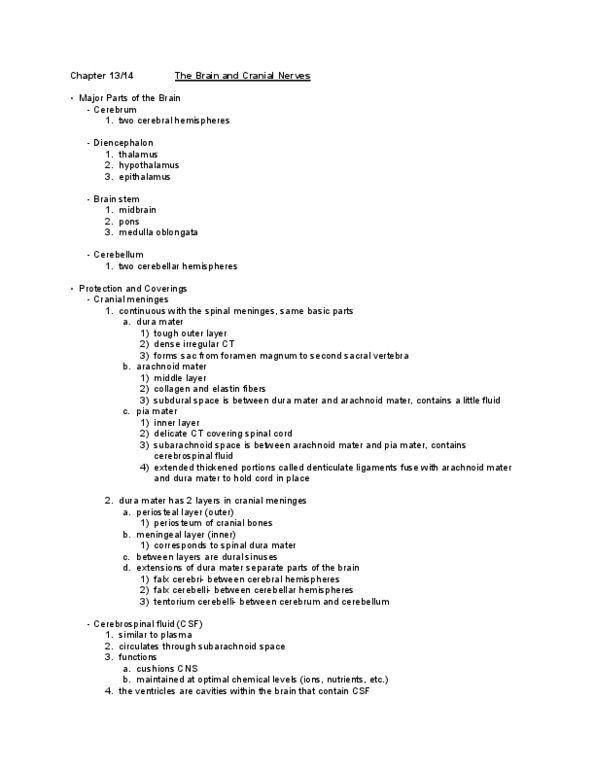
Dura mater: tough outer layer: sub-dural space: beneath dura. Arachnoid mater: thin, transparent middle layer: subarachnoid space: beneath arachnoid. Pia mater: transparent inner layer, outside covering of nerve tissue: adhere to brain and cord. Epidural space (above dura)- in cord only. 2 cups daily produced and reabsorbed circulates around brain and cord. Central sulcus: groove dividing frontal and parietal. Sensory & motor functions for opposite side of body. Left hemisphere (90% dominant: language, analytical reasoning. Right hemisphere (non-dominant: non-verbal functions (musical patterns/spatial relationships, imagination, artistic skill, higher intellectual processes, concentration, planning, complex problem solving. Temporal: meaning of sensory information, understanding speech, choosing words to express thoughts and feelings, understand speech, read printed words, remember visual scenes, and music, wernicke"s area- speech area . Speaking but cannot understand the words: analyze visual patterns. Mapping of motor/sensory cortexes inferior extremity: most medial superior extremity: middle superior/cranial: most lateral size represents degree of representation.