ANAT2111 Lecture Notes - Lecture 6: Internal Auditory Meatus, Tympanic Cavity, Outer Ear

21 Jun 2021
School
Department
Course
Professor
Document Summary
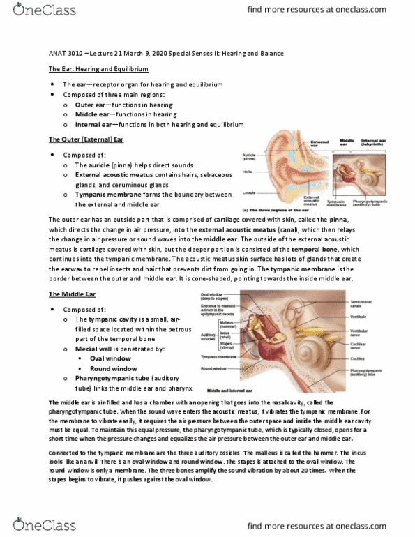
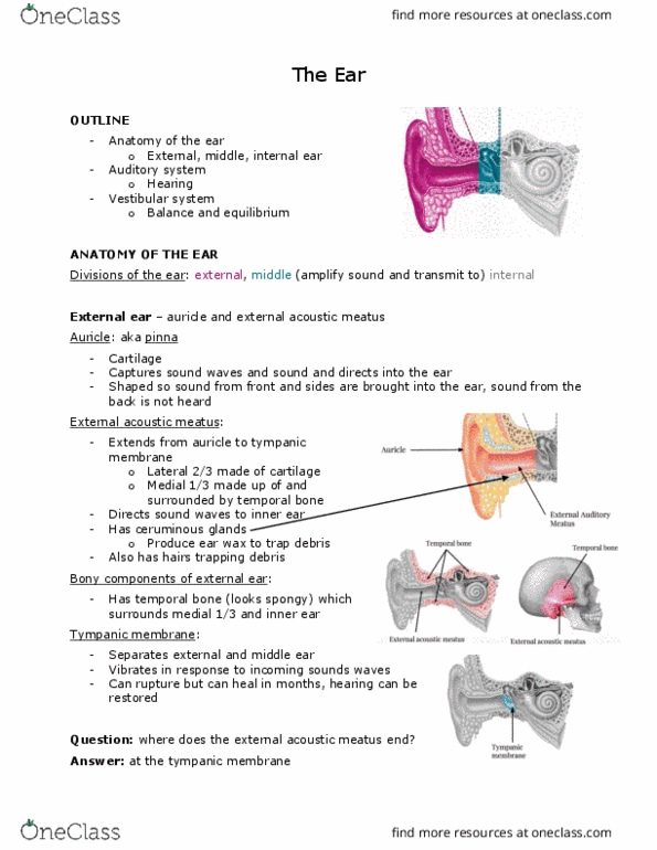
Located in the temporal bone internal and external acoustic meatus. External ear = first part of sound transduction. Middle ear = air filled cavity within petrous part of temporal bone. Bound laterally by middle ear and medially by internal acoustic meatus. Pinna (auricle) - helical shape - acts as a funnel that collects, localises and resonates sound into the ear canal. External acoustic meatus - pinna to tympanic membrane . Lateral 1/3 = cartilaginous + medial 2/3 = bony. Passage lined with ceruminous glands, sebaceous glands, hair follicles protect and clean the ear. Laterally covered by epidermis + medially mucous membrane. Vibrates according to pitch and wavelength, but moves 1/billionth of cm. Divided into epitympanic recess superiorly + tympanic cavity inferiorly. Transduces vibrations from tympanic membrane to internal ear via auditory ossicles (malleus, incus and stapes). Round window"s flexibility allows movement of stapes against oval window cause displacement of perilymph within the internal ear allowing vibration to enter the labyrinth.