ANHB2212 Final: ANHB2212 EXAM NOTES: Vertebral Column

3 Jun 2018
School
Course
Professor

Vertebral Column
Growth and Development of the Vertebral Column:
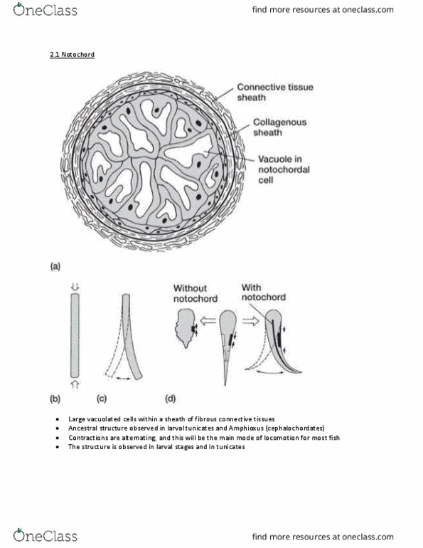
• Notochord induces segmentation
• Mesoderm either side condenses into blocks → somites
o Dermatome
o Myotome → vertebral muscle
o Sclerotome → vertebrae
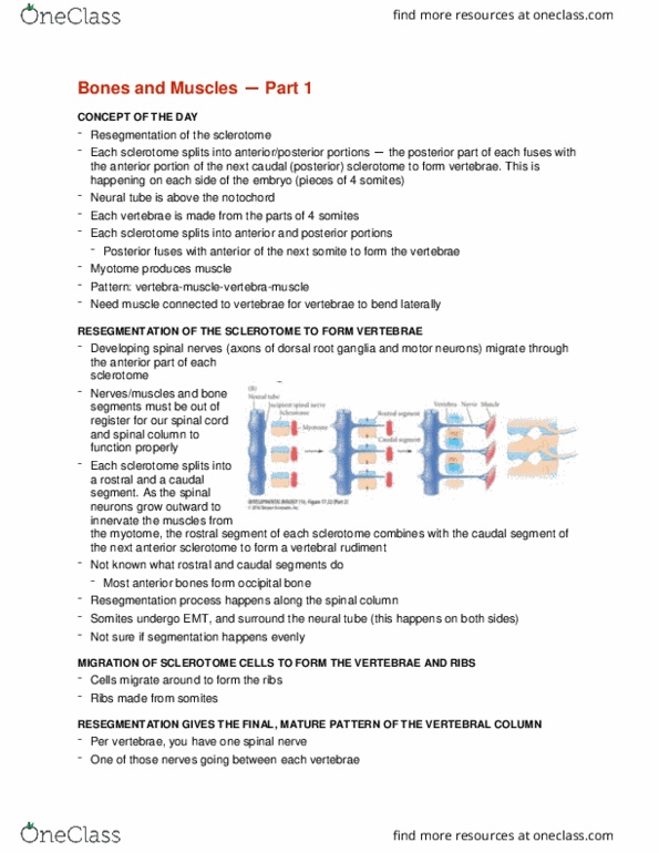
• Vertebrae are intersegmental
o Each vertebra forms from 2 adjacent sclerotomes
o Each muscle attaches to 2 vertebrae
• Sclerotome divides into cranial and caudal halves → caudal end of one
segment fuses with cranial end of segment behind it → resegmentation
• Body wall vessels lie between somites → intersegmental arteries
• Arteries provide better nutrition to perinotochordal cells
o Perinotochordal cells close to arteries become vertebral bodies
o Less nourished parts become intervertebral discs
o Notochord squeezed into zone between developing bodies
• 3 Stages of vertebral development
o Mesenchymous stage → weeks 4-6
o Cartilaginous stage → weeks 6-9
o Osseous stage → week 8 onwards
• Mesenchymous stage
o Endochondral ossification
o Sclerotome cells migrate to form
▪ Perinotochordal sheath (centrum)
▪ Neural arch
▪ Costal element
• Cartilaginous stage
o Begins with mesenchymous scaffold of vertebra
o Paired chondoficiation centers appear in
▪ Centrum
▪ Neural arch
▪ Costal element
find more resources at oneclass.com
find more resources at oneclass.com

o Mesenchyme gradually replaced by hyaline cartilage
o In regions where discs form, fibrocartilage forms ring (annulus
fibrosis) around notochordal element (nucleus pulposis)
▪ Discs lie in line with rest of somite
o Anomalities
▪ Because cartilage centers are paired, if one side fails to
form, vertebrae develop asymmetrically → hemivertebrae
• Osseous stage
o Begins with a cartilage model of the vertebrae
o Centers of ossification appear
▪ Centrum → unpaired
▪ Neural arches
▪ Costal elements → either fuse with the rest of vertebrae, or
become ribs and develop joints
o Bones grow but cartilage growth plates continue to separate
ossification centers
▪ Neurolaminar
▪ Neurocentral
o Anomalities → if notochord inhibits ossification
▪ Ossification of the centrum is inhibited if too many
notochordal cells remain between the vertebrae →
butterfly vertebrae
▪ Ossification can obliterate the disc if too few notochordal
cells remain between vertebrae → block vertebrae
find more resources at oneclass.com
find more resources at oneclass.com

• Parts of all adult vertebrae are derived from the centrum, neural arch and
costal elements
• Centrum doesnt make the whole vertebral body → neural arches
contribute laterally
• Costal elements form ribs in thoracic region, but in other regions it is
incorporated into parts of the transverse process
• Post-natal growth of vertebral column
o Vertebral canal stays about the same size from birth, but
surrounding bone must grow larger
o At birth, centrum and neural arch elements still separate by
cartilate growth plates
▪ Interlaminar
▪ Neurocentral
o By 6-8 years old
▪ Growth plates close
▪ Endplates and processes are unossified
▪ Vertebrae cant increase diameter but can increase height
o By puberty
▪ Secondary ossification centers appear
• Tip of spinous process
• Tip of transverse process
• Ring apophysis
▪ Additional growth in height is due to ring apophysis
o By adulthood
▪ All epiphysis close
▪ Growth can only continue by surface remodelling
Regional Evolution of the Vertebral Column:
• Over course of evolutionary history the vertebral column has become
more regionally specified and adapted for
o Environment → aquatic vs. terrestrial
o Locomotive strategies → quadrupedal, bipedal, arboreal
• Fish
o Only have thoracic and caudal vertebrae
o Vertebrae cranial to the cloaca have ribs
o All vertebrae caudal to cloaca are tail vertebrae
o No neck → no need to position head
o Lateral septum → transverse process in other vertebrates
• Amphibians
o Have hind limbs → sacrum
o Sacrum attaches hind limbs to axial skeleton
o Efficient transfer of force from hind limb to body
o No neck → specializations for obtaining food
find more resources at oneclass.com
find more resources at oneclass.com
Document Summary
Intervertebral foramina: vertebral canal is larger where there is more potential for movement lumbar and cervical spinal cord also larger, vertebral canal is smallest in the thorax and the sacrum. Join the tip of adjacent spinous processes: strong band of white fibrous tissue drawn taught by full flexion, indistinct below l4 lumbar fascaia is thick, replaced by ligamentum nuchae in neck. Interspinous ligament: unite spinous processes along adjacent borders, relatively weak bands of fibrous tissue, longest and strongest in lumbar region fuse with supraspinous ligament. Intertransverse ligament: unite transverse processes on adjacent borders, similar sheets of weak fibers, generally absent in cervical region, recognizable in lumbar region, facet joints. Joint capsules are relatively lax in cervical region. Intrinsic postvertebral muscles: unisegmental intertransveral system, deep layer of the extensor muscles of the back, generally all supplied by dorsal rami of spinal nerve, intertransverseraii.