PSYCH 1XX3 Study Guide - Final Guide: Radioactive Tracer, Wilder Penfield, Inferior Colliculus

20 Feb 2017
School
Department
Course
Professor
Document Summary
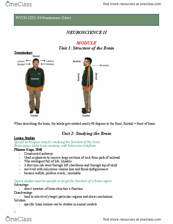
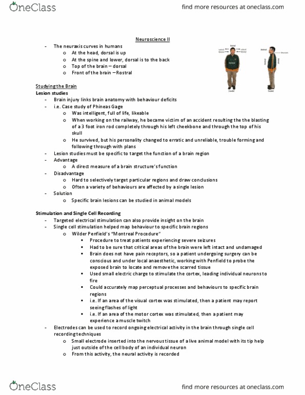
Rostral the front of the brain. Caudal the back of the brain. Ventral the bottom of the brain. Dorsal the top of the brain. Special techniques exist for studying the structure and function of the brain: comes from work with animals, tissues and special preparations. Brain injury links brain anatomy with behavioural deficit: phineas gage 1848. Using explosives to move ground for railways. Iron rod pierces through left cheekbone and top of skull. Often a variety of behaviours are affected due to the lesion. Single cell stimulation helped map behaviour to specific brain regions. Reveals function of individual neurons: cats presented with visual stimuli while recording single cells, cells responded to specific categories of stimuli. Large scale structures are studied using neuroimaging techniques. Ct: produces structural slices of the brain, helpful to diagnose brain injuries, low resolution. Mri: higher resolution image, magnetic fields generated which align hydrogen atoms in the brain, localizes tissue precisely.