[Kinesiology 3336A/B] - Final Exam Guide - Everything you need to know! (38 pages long)

29 Nov 2016
School
Department
Course
Professor
Document Summary
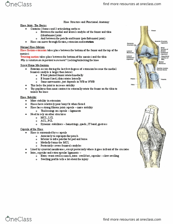
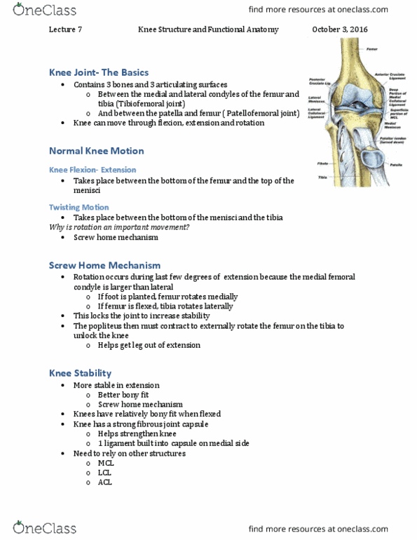
Stability comes from: shape of bones (mortise, capsule & ligaments, strength of muscles (dynamic stabilizers) 85% lateral, 10% syndesmosis (high ankle injury), 5% medial. Made up of: lower end of tibia, medial malleolus, lateral malleolus, transverse tib/fib ligament. Lateral malleolus longer and more posterior than medial: adds significant stability. Trochlear surface is wider anteriorly than posteriorly. Has no muscles that attach to it. With dorsiflexion the wider portion lies between the malleoli: better bony fit, more stable. Thin & weak anteriorly & posteriorly to allow movement. Atfl- weakest of all 3 lateral ligaments (no bony fit: strain increases with pf/ inversion. Cfl- extra-capsular (less swelling than capsular: stabilizes subtalar joint and limits adduction/ medial tilt, tight in neutral dorsiflexion. Ptfl- strongest of all 3 lateral ligaments (dorsiflexion: strong in dorsiflexion b/c large anterior surface on talus. September 13, 2016: limits talar abduction or lateral tilt. Injury can be closely correlated to the load-deformation curve.