MEDRADSC 2D03 Chapter Notes - Chapter 1: Pleural Cavity, Hounsfield Scale, Voxel

21 Sep 2016
School
Department
Course
Professor
Document Summary
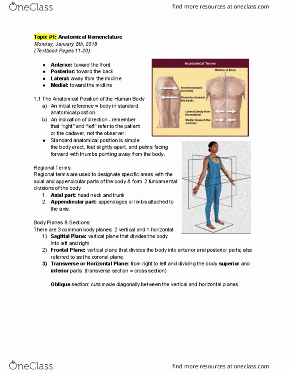
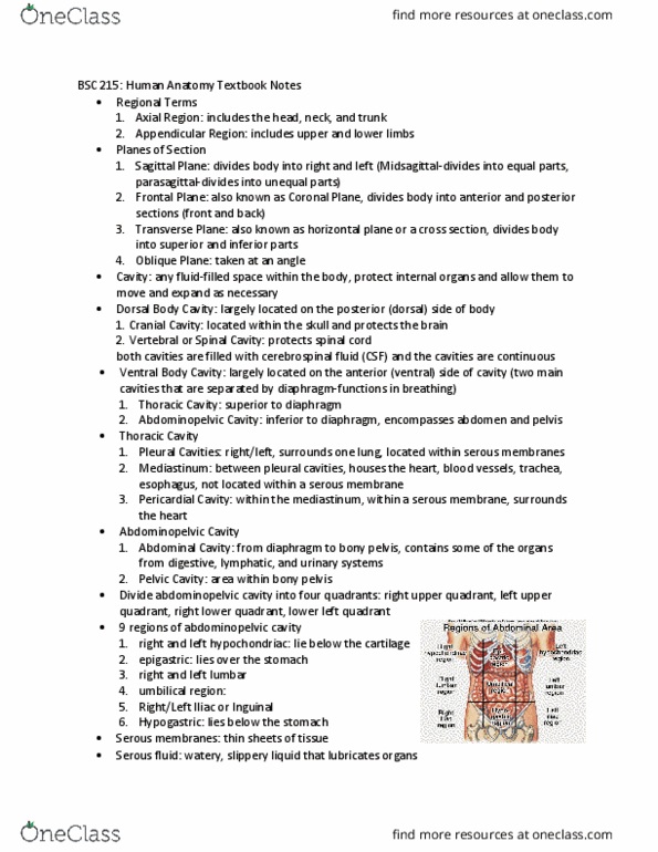
Sectional anatomy emphasizes the physical relationship between internal structures. Sagittal: vertical, dividing body into left and right. Coronal: vertical, dividing body into front and back. Oblique: passes diagonally between the axes of two other planes. Ventral- thoracic (two lateral pleural cavities and mediastinum) and abdominopelvic (abdominal and pelvic) cavities. Abdomen bordered superiorly by the diaphragm and inferiorly by the superior pelvic aperture. Each digital image can be divided into individual regions called pixels or voxels that are then assigned a numerical value corresponding to a specific tissue property of the structure being imaged. The numerical value of each voxel is assigned a shade of gray for image display. In ct, the numerical value (ct#) is referenced to a hounsfield unit (hu), which represents the attenuating properties or density of each tissue. Water is used as the reference tissue and is given a value of 0.