SAR HS 369 Chapter Notes - Chapter 4: Costal Cartilage, Thoracic Vertebrae, Thoracic Inlet

2 Jul 2020
School
Department
Course
Professor
Document Summary
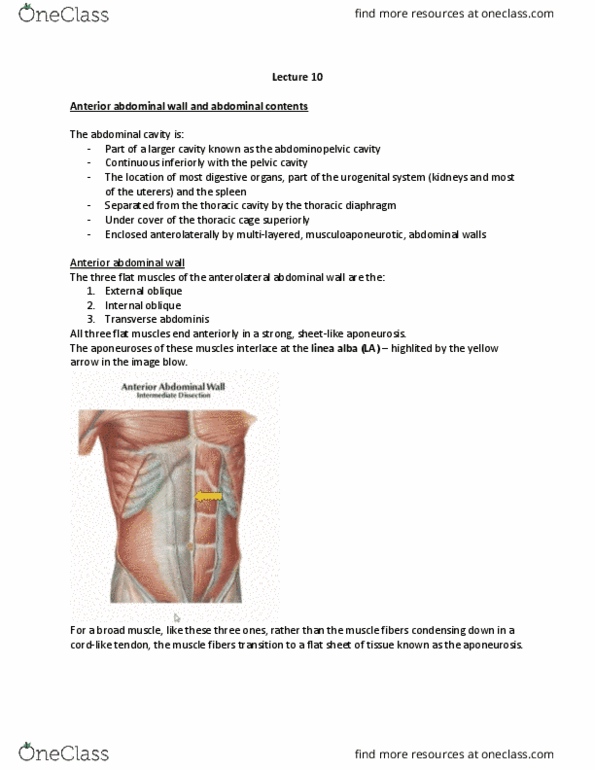
The thoracic wall consists of skin, fascia, nerves, vessels, muscles, cartilages and bones. Resisting the negative internal pressures generated by the elastic recoil of the lungs and inspiratory movements. Providing attachment for and supporting the weight of the upper limbs. Providing attachment for many of the muscles of the upper limbs, neck, abdomen and back and muscles of respiration. The thoracic skeleton forms the osteocartilaginous thoracic cage. 12 pairs of ribs and costal cartilages: costal cartilages form the anterior continuation of the ribs, providing a flexible attachment to the sternum. 12 thoracic vertebrae and intervertebral iv disks. The ribs are separated by intercostal spaces, which are occupied by intercostal muscles, vessels and nerves. The thoracic cavity communicates with the neck and upper limb through the superior thoracic aperture (thoracic inlet). Structures that go through the aperture are: trachea, esophagus, vessels and nerves. Because of the obliquity of the first pair of ribs, the superior thoracic aperture slopes antero- inferiorly.