MEDS12002 Lecture Notes - Lecture 4: Hepatomegaly, Wound, Phosphatase


1 | P a g e
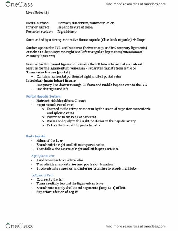
The liver – week 4
The liver
• Occupies the right hypochondrium, epigastrium and left
hypochondrium
• Measures 15 – 17cm
(Mid Clavicular Line)
• Weighs approx 1500g
• Covered by Glisson’s Capsule
• In epigastrium, liver extends several centimetres below the
xiphoid process
The four lobes
• Right Lobe
• Left Lobe
• Caudate Lobe
• Quadrate Lobe
Left Lobe:
• Medial and lateral segments by left hepatic vein & ligamentum teres
• Seperated from caudate lobe by ligamentum venosum
• Seperated from right lobe by middle hepatic vein superiorly and main
• lobar fissure inferiorly
Right Lobe:
• Six times larger than left lobe
find more resources at oneclass.com
find more resources at oneclass.com

2 | P a g e
• Divided into anterior and posterior segments by right
hepatic vein
• 3 posterior fossa: gallbladder, porta hepatis & inferior
vena cava
Caudate Lobe:
• Smallest lobe of liver
• Seperated from left lobe by ligamentum venosum
• Arterial supply through right and left portal veins and hepatic arteries
Quadrate Lobe:
• Situated on inferior visceral surface of liver
• Bounded posteriorly by porta hepatis and laterally by gallbladder fossa
Liver segments
The liver is attached to the diaphragm, anterior abdominal wall, stomach and retroperitoneum by
ligaments:
• Coronary Ligament
• Falciform Ligament
• Ligamentum Teres (Round Ligament)
• Gastrohepatic Ligament
• Hepatoduodenal Ligament
• Right & Left Triangular Ligaments
• Ligamentum Venosum
find more resources at oneclass.com
find more resources at oneclass.com

3 | P a g e
Coronary and triangular ligaments
The Coronary Ligament
• Connects posterior liver to diaphragm
• Upper layer is peritoneum from upper margin of the
bare area to the undersurface of diaphragm
• Lower layer is lower margin of bare area to the right
kidney, also known as hepatorenal ligament
Right & Left Triangular Ligaments
• Most lateral portion of the coronary ligament
• Connects liver to the body wall
Falciform ligament and Ligamentum Teres
Falciform Ligament
• Attaches the liver to the anterior abdominal wall
• Extends from diaphragm to the umbilicus
• Seperates the right and left subphrenic space
Ligamentum Teres
• Lies within the falciform ligament
• Previous fetal umbilical vein
Fetal circulation
• The Umbilical Vein carries oxygenated blood from
the placenta to the fetus & bypasses the liver via the
Ductus Venosus
• After birth both veins close and become ligaments
• The Umbilical Vein becomes the Ligamentum Teres
• The Ductus Venosus becomes the
Ligamentum Venosum
Ductus venosus and Umbilical Vein
find more resources at oneclass.com
find more resources at oneclass.com
Document Summary
The liver: occupies the right hypochondrium, epigastrium and left hypochondrium, measures 15 17cm (mid clavicular line, weighs approx 1500g, covered by glisson"s capsule. In epigastrium, liver extends several centimetres below the xiphoid process. Left lobe: right lobe, caudate lobe, quadrate lobe. Left lobe: medial and lateral segments by left hepatic vein & ligamentum teres, seperated from caudate lobe by ligamentum venosum, seperated from right lobe by middle hepatic vein superiorly and main lobar fissure inferiorly. Right lobe: six times larger than left lobe. 1 | p a g e: divided into anterior and posterior segments by right hepatic vein, 3 posterior fossa: gallbladder, porta hepatis & inferior vena cava. Caudate lobe: smallest lobe of liver, seperated from left lobe by ligamentum venosum, arterial supply through right and left portal veins and hepatic arteries. Quadrate lobe: situated on inferior visceral surface of liver, bounded posteriorly by porta hepatis and laterally by gallbladder fossa.