BIOL123 Lecture Notes - Lecture 4: Phase-Contrast Microscopy, Polarizer, Photomultiplier


Lecture 4: Microscopy instruments and applications
1
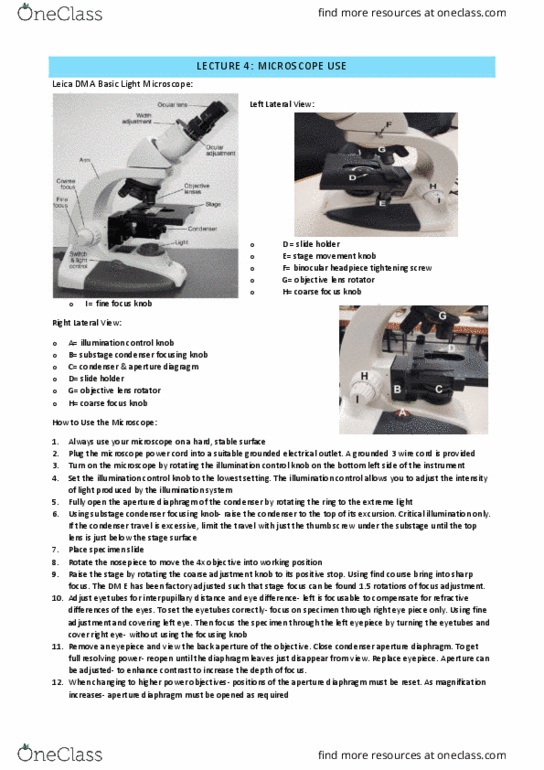
Microscope Focusing
1. Start with the lowest objective (x4) and make sure the condenser is close as possible to the stage
2. Adjust the width of the eyepieces on the binocular scope until you see one circle of light in view
3. Add slide with specimen to stage and make sure firmly in place
4. Check to see fine focus is about mid-range in its range of movement
5. Use coarse adjustment to get specimen in focus
6. Fine focus can then be used to sharpen the focus
7. Use iris diaphragm to adjust light intensity (controls aperture)
8. Move to next objective (x10) and adjust diaphragm to increase light
9. Move to next objective (x40) and adjust diaphragm again, use fine focus
10. The highest one usually needs immersion oil to view
Optical Light Microscope
• Image magnified by focusing light with glass lenses on a thin
tissue section
• Brightfield illumination used
o White light is transmitted through the dense areas of
the sample
• Specimens stained with coloured dyes
• Resolution of 0.2 μm

Lecture 4: Microscopy instruments and applications
2
Microscopy
PHASE CONTRAST MICROSCOPY
• Contrast enhancing technique
• Helps to view transparent cells and tissues – allows living cells and ongoing biological processes to be
observed and recorded
o Staining effects biological function
POLARISED LIGHT MICROSCOPY
• Used to analyse structures that have two different xive indices
• Provides info on the orientation of molecular structures in a specimen
• Polariser positioned in light path somewhere before the specimen
• Analyser (2nd polariser) placed after the objective

Lecture 4: Microscopy instruments and applications
3
FLORESCENCE MICROSCOPY
• Stained with multi coloured fluorescent dyes that are visible under UV light
• Used to observe distribution of biological materials in cells and tissues
(proteins, lipid and nucleic acids)
• Sensitivity of fluorophores means lower concentrations of biological
material can be detected than with conventional dyes
CONFOCAL MICROSCOPY
• Controllable depth of field
• Elimination of out of focus information
• Ability to collect serial optical sections from thick specimens
• Used with fluorescence microscopy
• Image captured by photomultiplier
TRANSMISSION ELECTRON MICROSCOPY
• Measures relative differences in transparency (contrast) of a
specimen to electrons
• Ultrathin tissue sections are stained with heavy metals to increase
contrast
• Specialised labelling for structures
• Images are coloured and a resolution of 0.2 to 0.5 nm
SCANNING ELECTRON MICROSCOPY (SEM)
• Produces images of 3D surfaces
• Electron bean is scanned across a specimen and the emission of electrons is measured from
specimens coated with a thin film of metal (such as platinum)
• Images are colourless have a resolution of ~2.5 nm
ATOMIC FORCE MICROSCOPE
• Probe a specimen surface with an ultra-sharp tip reconstructing electronically an image with
nanometre resolution
• “Atomic force” means they are so sensitive they actually can record forces as small as the
interactions between individual atoms on the surface and at the tip
• Different sizes, shapes, stiffnesses, resonant vibration frequencies, conductivities, optical, magnetic,
or chemical properties, to enable the many operating modes already developed for such microscopes
Document Summary
Image magnified by focusing light with glass lenses on a thin tissue section: brightfield illumination used, white light is transmitted through the dense areas of the sample, specimens stained with coloured dyes, resolution of 0. 2 (cid:373) Images are coloured and a resolution of 0. 2 to 0. 5 nm. Images are colourless have a resolution of ~2. 5 nm. B r i g h t f i el d i l l u m i n a t i o n. C r i t i c a l i l l u m i n a t i o n: uneven illumination causes artefacts to shadow and glare to image, results in bulb filament in image. Light source falls in the same plane as the image of the specimen. Light sources: consider emission characteristics of source. Light-gathering power of the objective (governs image brightness) L i g h t p a t hw a y s.