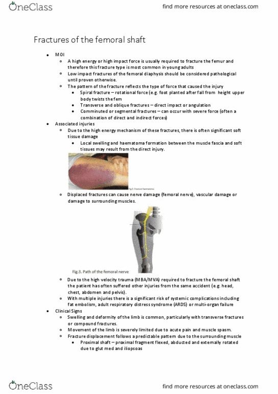
PHTY206 Lecture Notes - Lecture 20: Neurovascular Bundle, Common Peroneal Nerve, External Fixation


Fractures of the knee
• Supracondylar fractures
o Classification and mechanism of injury (MOI)
• A supracondylar fracture involves the distal femur and is usually non-articular,
however it can extend into the epiphysis and indicate an intra-articular
fracture.
• There are several proposed classification systems however the AO
classification is the most comprehensive
▪ Divides the fractures into:
• Extra-articular (Type A)
• Unicondylar (Type B)
• Bicondylar (Type C)
▪ With these categories then further subdivided into depending on their
severity.
• Mechanism of Injury
▪ Occur in both the elderly and young patients but with very different
mechanisms of injury.
▪ In young patients the mechanism is usually high energy
• E.g. impact against a dashboard with the knee flexed during a
motor vehicle accident.
▪ In more elderly patients the mechanism is often low energy
• E.g. a fall onto the flexed knee with bone already weakened by
osteopaenia.
▪ Will often be displaced with the distal fragment posteriorly angulated
due to the pull of the gastrocnemius muscle.
• Associated Injuries
▪ Due to the high energy MOI there are often associated injuries with
supracondylar fractures.
▪ This can include both remote life threatening injuries (head, chest,
major vascular) and local injuries.
o Orthopaedic management - surgical
• Open reduction and internal fixation is the management of choice for
supracondylar fractures.
• Surgical management is focused on anatomical reconstruction of the articular
surface and realignment in all axes of the metadiaphyseal segment to avoid
deformities.
• Small residual deformities may increase the risk of, or accelerate
degenerative changes in the articular cartilage of the knee joint.
find more resources at oneclass.com
find more resources at oneclass.com

o Orthopaedic management - conservative
• If a patient is medically unstable or the supracondylar fracture is severely
comminuted surgery may not be a viable option and skeletal traction can be
considered.
• This conservative method of treatment is associated with a high risk of
malunion including varus/valgus and rotational deformities due to the pull of
the gastrocnemius muscle.
• There are also a range of potential complications of skeletal traction due to
pin sites and prolonged bed rest.
• Casting is another conservative management option, particularly for
extracapsular fractures but the risk of malunion remains high
o Physiotherapy management
• Following ORIF should include:
▪ Daily circulo-respiratory assessment and maintenance exercises
▪ Strengthening exercises to focus on regaining quadriceps control (static
quads, IRQ, SLR).
▪ Gentle range of movement exercises at the knee are important if the
fracture fixation is stable and this will be guided by the surgeon.
▪ Mobilisation should commence day 1, initially on a rollator and
progressing quickly to crutches
▪ The weight bearing status will be restricted from NWB to TWB for
several weeks until the surgeon gives permission for weight bearing to
be progressed.
• Following skeletal traction
▪ Physiotherapy management will involve gradual protected mobilisation
and ROM exercises
▪ Extreme care however should be taken when commencing ROM of the
knee joint ensuring that the frature is stale and won’t e displaed
with movement (approximately 4 weeks)
▪ The split bed technique may be used for knee ROM and IRQ exercises.
• Older patients following conservative or surgical management who may not
cope NWB may be mobilised in a cast brace to allow earlier weight bearing
o Complications
• Early Complications
▪ Arterial damage – small but definite risk
▪ Common in the elderly therefore often secondary complications of
myocardial infarctions, chest infections and deep wound infection
find more resources at oneclass.com
find more resources at oneclass.com

• Late Complications
▪ Joint stiffness – related to scarring during surgery and from the injury
itself.
• Physiotherapy must focus on knee joint ROM
▪ It is unlikely full ROM will be regained
▪ Muscle Weakness
▪ Malunion – posterior angulations or varus deformities are common
▪ Non-union
• Patella fractures
o Classification and MOI
• Patella fractures are usually described as non-displaced or displaced.
• Displaced fractures involve a step in the articular surface or more than 3mm
of fragment separation.
• Patella fractures can also be described according to the pattern of fracture
line as transverse, longitudinal, polar or comminuted (stellate).
• MOI
▪ Most patellar fractures result from a direct blow or fall onto the knee
•
With these fractures the extensor mechanism usually remains
intact, however there is frequently damage to the articular
surface
•
This may result in an undisplaced crack or a comminuted (stellate)
fracture.
▪ A forceful contraction of the quadriceps may also cause a transverse
fracture of the patella, and if displaced this can result in a retinacular
tear and extensor mechanism deficit.
▪ Bipartite patella is a common congenital fragmentation of the patella
occurring in approximately 1% of the population
•
Care should always be taken not to mistake a bipartite patella for a
patella fracture – a bipartite patella should have a smooth regular
line and is often bilateral.
find more resources at oneclass.com
find more resources at oneclass.com
Document Summary
In young patients the mechanism is usually high energy: e. g. impact against a dashboard with the knee flexed during a motor vehicle accident. Surgical management is focused on anatomical reconstruction of the articular surface and realignment in all axes of the metadiaphyseal segment to avoid deformities. Small residual deformities may increase the risk of, or accelerate degenerative changes in the articular cartilage of the knee joint: orthopaedic management - conservative. Following orif should include: daily circulo-respiratory assessment and maintenance exercises. Joint stiffness related to scarring during surgery and from the injury itself: physiotherapy must focus on knee joint rom. Indicated for undisplaced fractures with an intact articular surface: treatment involves either a cylinder cast or a knee immobiliser such as a. K-wires and tightened over the front of the patella. Facilitation techniques may be used to strengthen vmo, and patellofemoral mobilisation techniques may help restore normal patellofemoral tracking: mobilisation can commence immediately in the brace and fwb is allowed.