PHTY206 Lecture Notes - Lecture 12: Synovitis, Neuroma, Pes Cavus


Physical examination of the foot
• Discuss common acute injuries of the foot
• Describe the mechanism of injury of the ligaments of the foot
• Describe common overuse injuries which affect the tissues of the foot
• Describe the common changes in foot and ankle biomechanics which may lead to the
development of tissue injury in the foot
• Differentiate injured structures and/or pathology of the foot using signs and symptoms
from the patient interview and physical examination
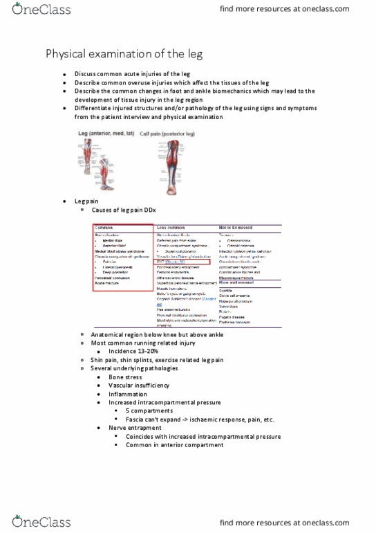
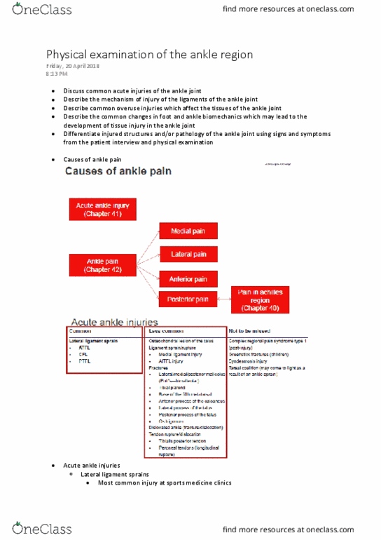
• Causes of rearfoot pain
o Plantar heel pain
• Most common cause of heel pain
• Overuse condition of plantar fascia and its attachment to calcaneus
• Resembles tendinopathy
▪ Due to collagen disarray
▪ Absence of inflammatory cells
• Evidence of predisposing factors
▪ Limited ankle DF ROM
▪ High BMI
▪ Running (volume/repetitive stress)
▪ Work related WB activities
• Patient interview
▪ Onset
• Gradual, insidious
▪ Pain
• Medial aspect of heel
▪ Behaviour
• Worse a.m. "first step pain" -> also if sitting/inactive for a while
then first get up
• Initially improves with activity
• Ache post-activity
▪ Progression
find more resources at oneclass.com
find more resources at oneclass.com

• As condition becomes more severe pain with WB, worsens with
activity, NWB pain
• Physical exam
▪ Acute tenderness medial tuberosity of calcaneus +/- along plantar fascia
▪ +ve Windlass (Jack's) test
• Passive DF first MTP joint +/- palpation provokes pain
▪ Predisposing factors (ankle DF)
• Imaging can be useful
▪ Ultrasound: swelling and thickness (>4mm)
▪ X-ray: spur
▪ MRI: thickening, bone marrow, oedema, spur
o Fat pad contusion
• Fat pad composed of elastic fibrous tissue septa separating closely packed fat
cells
• Acts as shock absorber
• Protects calcaneus
• Causes
▪ Fall onto heel from height
▪ But occasionally chronic due to excessive heel strike with poor
cushioning
• Patient interview
▪ Marked lateral heel pain
▪ Increase with WB
▪ Esp. heel contact
▪ Hx traumatic event (usually)
find more resources at oneclass.com
find more resources at oneclass.com

• Physical exam
▪ Tenderness posterolateral heel
▪ May be redness
▪ MRI: oedematous changes
• Less common causes of rearfoot pain
o Calcaneal stress #
• 2nd most common tarsal stress fracture
• Either upper posterior margin or adjacent to medial tuberosity
• Esp. military (marching) runners, ballet, dancers, jumping athletes
• Patient interview
▪ Insidious onset heel pain
▪ Aggravated by WB
• Physical exam
▪ Localised tenderness
▪ Pain reproduced by squeezing posterior aspect of calcaneus from both
sides simultaneously
o Medial calcaneal nerve entrapment
• Branch of posterior tibial nerve arising at level of medial malleolus
• Innervates skin of heel
• Patient interview
▪ Burning pain of inferomedial aspect of calcaneus - often radiates to arch
of foot
▪ Aggravated by running
• Physical examination
find more resources at oneclass.com
find more resources at oneclass.com
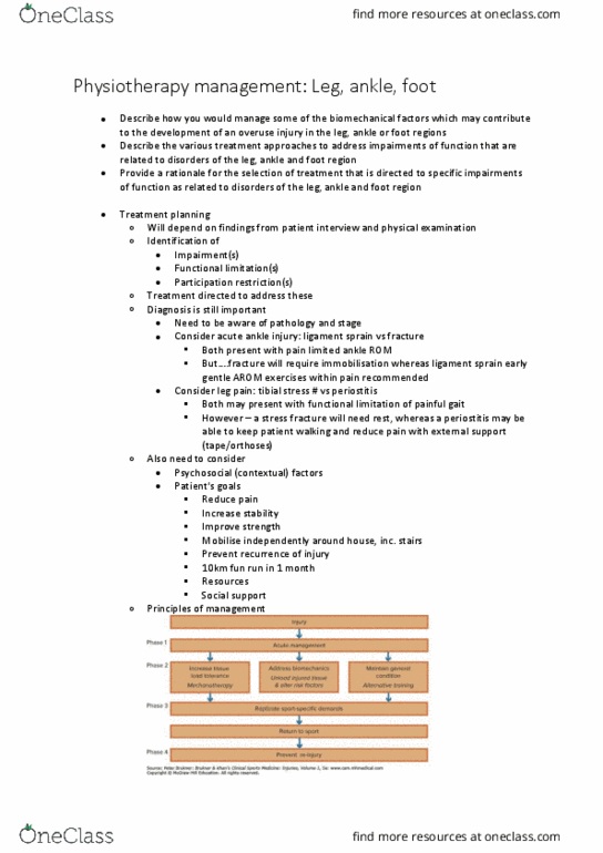
Document Summary
Imaging can be useful: ultrasound: swelling and thickness (>4mm, x-ray: spur, mri: thickening, bone marrow, oedema, spur, fat pad contusion. Fat pad composed of elastic fibrous tissue septa separating closely packed fat cells: acts as shock absorber, protects calcaneus, causes. Fall onto heel from height: but occasionally chronic due to excessive heel strike with poor cushioning, patient interview, marked lateral heel pain. Increase with wb: esp. heel contact, hx traumatic event (usually, physical exam, tenderness posterolateral heel, may be redness, mri: oedematous changes. Less common causes of rearfoot pain: calcaneal stress , 2nd most common tarsal stress fracture, either upper posterior margin or adjacent to medial tuberosity, esp. military (marching) runners, ballet, dancers, jumping athletes, patient interview. Insidious onset heel pain: aggravated by wb, physical exam. Localised tenderness: pain reproduced by squeezing posterior aspect of calcaneus from both sides simultaneously, medial calcaneal nerve entrapment, branch of posterior tibial nerve arising at level of medial malleolus, patient interview.