PHTY206 Lecture Notes - Lecture 11: Ganglion, Idiopathy, Nerve Conduction Study


Physical examination of the ankle region
Friday, 20 April 2018
8:13 PM
• Discuss common acute injuries of the ankle joint
• Describe the mechanism of injury of the ligaments of the ankle joint
• Describe common overuse injuries which affect the tissues of the ankle joint
• Describe the common changes in foot and ankle biomechanics which may lead to the
development of tissue injury in the ankle joint
• Differentiate injured structures and/or pathology of the ankle joint using signs and symptoms
from the patient interview and physical examination
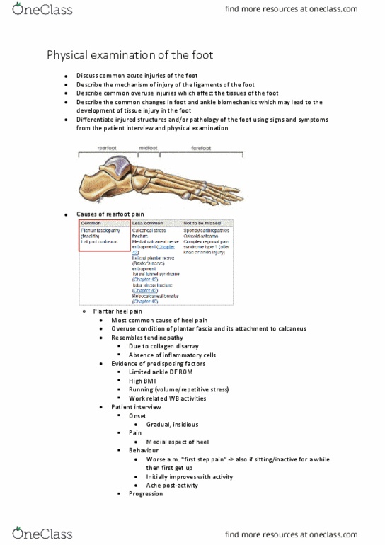
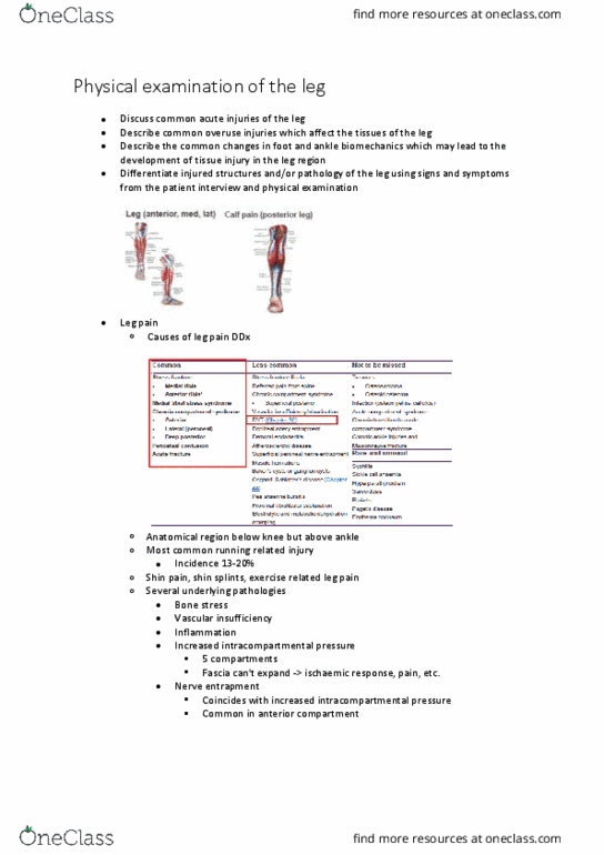
• Causes of ankle pain
• Acute ankle injuries
o Lateral ligament sprains
• Most common injury at sports medicine clinics
find more resources at oneclass.com
find more resources at oneclass.com

• High incidence in football, basketball, netball
• 80% ankle sprains caused by inversion/supination
• 4x more inversion injuries
▪ Relative instability of lateral joint
▪ Relative weakness lateral ligaments
• MOI
▪ INV suggests lateral lig, EVN suggests medial ligs
▪ Compressive forces consider osteochondral injury
▪ Sometimes audible snap/crack/tear
• Onset
▪ Able to WB immediately suggests sprain
▪ Unable to WB suspect #
• Location
▪ Pain/swelling/bruising
• Gives indication of ligament involved
• Stress tests
▪ More accurate few days post injury
• Diagnostic imaging
o Lateral ligament sprains - ATFL
• First ligament to tear - torn 97% of cases
• MOI
▪ PF and INV
• Physical exam
▪ Localised pain (antero-lateral)
▪ Localised swelling (antero-lateral)
▪ +ve anterior drawer
find more resources at oneclass.com
find more resources at oneclass.com

o Lateral ligament sprain - CFL
• Usually in conjunction with ATFL
▪ Isolated rupture rare (3% of inversion injuries)
• Physical exam
▪ Localised pain (lateral, below lateral malleolus)
▪ +ve talar tilt test (INV)
o Lateral ligament sprain - PTFL
• Last ligament to tear
• Complete tear of ATFL, CFL and PTFL rare and results in dislocation of ankle joint
• Isolated rupture of PTFL rare
find more resources at oneclass.com
find more resources at oneclass.com
Document Summary
Inv suggests lateral lig, evn suggests medial ligs. Sometimes audible snap/crack/tear: onset, able to wb immediately suggests sprain, unable to wb suspect # Location: pain/swelling/bruising, gives indication of ligament involved. Stress tests: more accurate few days post injury, diagnostic imaging, lateral ligament sprains - atfl. First ligament to tear - torn 97% of cases: moi, pf and inv, physical exam. Localised swelling (antero-lateral: +ve anterior drawer, lateral ligament sprain - cfl, usually in conjunction with atfl. Isolated rupture rare (3% of inversion injuries: physical exam. Localised pain (lateral, below lateral malleolus: +ve talar tilt test (inv, lateral ligament sprain - ptfl. Last ligament to tear: complete tear of atfl, cfl and ptfl rare and results in dislocation of ankle joint. Less common as medial ligament stronger: moi. Forced eversion: physical exam, medial pain on palpation and swelling, talar tilt test (evn)