MEDS12003 Lecture Notes - Lecture 5: Tunica Vaginalis, Deep Inguinal Ring, Mediastinum Testis


1 | P a g e
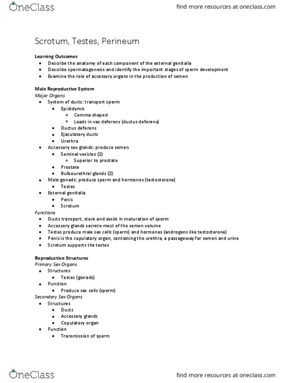
Week 5: Scrotum/testes
Anatomy scrotum
• Superficial facia and dartos muscle of the
scrotum form an incomplete scrotal septum
dividing the scrotum into Lt and Rt halves
▪ Septal raphe
Anatomy – Testes
• Paired ovoid glands measuring between 3-5cm
in length and 2-3cm in width and AP
measurement
• Size and weight decreases with increasing age
• Testes covered by tunica vaginalis
▪ 2 layers –parietal (outer), visceral
(inner)
find more resources at oneclass.com
find more resources at oneclass.com

2 | P a g e
• Tunica albuginea – a dense, white fibrous capsule
▪ Fold on tunica albuginea forms mediastinum testis
• Multiple, thin septa from the inner aspect of the tunica albuginea join posteriorly to form the
mediastinum testis.
Tunica Albuginea
Anatomy – Testes
Normal anatomy
• Mediastinum testis provides support for blood vessel and ducts
• The septa separate the testis into many lobules (> 200)
• Each lobule contains seminiferous tubules
• Seminiferous tubules join together into larger ducts which lead to the rete testis (located in the
mediastinum testis)
• Drains into the epididymis via 10-15 efferent ductules
Vasculature
• Blood supplied to testis via deferential, cremasteric and testicular arteries
• Venous drainage via pampiniform plexus
• Testicular venous drainage is important for investigation of varicoceles
▪ LT testicular vein joins LT Renal Vein but with a 90 degree configuration
▪ RT testicular vein drains into IVC
find more resources at oneclass.com
find more resources at oneclass.com

3 | P a g e
Anatomy – Epididymis
• Curved structure measuring
approx 6-7 cm
• Rete testis form into ductus
epididymis
• Consists of head, body and tail
• Head located at superior aspect of
the testis
• Tail loosely connected to inferior
aspect of the testis
Embryology
• The scrotal sac forms in the fourth month of
gestation
• The testes descend from their position at the 10th
thoracic level near the kidneys
• Descent dependent on gubernaculum-shortening
• Testes descend through the inguinal canal
• “Processus vaginalis” forms -8th week.
• 12th week –testes descend into deep inguinal ring
• Final descent occurs 7-9 months, or approx. around birth.
• Abnormalities may occur resulting in ectopic testes
▪ Higher risk of malignancy and infertility
Normal variants
• Embryologic remnants
▪ Appendix testis
o Mainly seen with hydrocele
▪ Appendix of epididymis
o Situated on epididymal head
Scrotum
contents
find more resources at oneclass.com
find more resources at oneclass.com