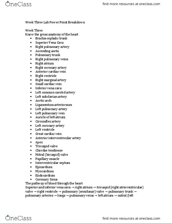
314151 Lecture Notes - Lecture 5: Mitral Valve, Interventricular Septum, Breathing

24 May 2018
School
Department
Course
Professor

Lecture 5 – 5/4/16
Function of the cardiorespiratory system:
- Acquisition – respiratory system including pulmonary ventilation, lungs, alveoli & gas diffusion.
- Circulation – cardiovascular system (heart, arteries, venous system)
- Transport – Blood including plasma, haemoglobin, platelets & leukocytes.
The role of the circulatory system is to transport and distribute key nutrients to tissues and remove any by-
products. Systemic circulation transports blood from the left ventricle of the heart to the body tissues.
Pulmonary circulation pumps blood from the right ventricle to the lungs.
A. Pulmonary artery – deoxygenated blood to the lungs
B. Pulmonary semilunar valve
C. Left atrium
D. Mitral valve
E. Aortic valve
F. Chorde tendone
G. Pupillary muscles
H. Left ventricle
I. Interventricular septum
J. Myocardium
K. Apex of the heart
L. Tricuspord valve
M. Right Atrium
N. Aorta – distributes blood to tissues
Cardiac Muscle – striated, binucleated. Sarcomeres of the heart are
joined end to end by intercalated discs made of Desmosomes (that
mechanically hold the cells together) and Gap Junctions (which, permit action potentials to propagate from
cell to cell).
Cardiac Cycle – all mechanical and electrical events that occur during one heartbeat.
Systole – Contraction phase – chambers eject blood, high pressure; ventricle twists, shortens and thickens.
Diastole – Relaxation phase – chambers fill with blood,
twice as long as systole. Ventricle untwists & lengthens.
Electrical activity of the heart:
Sinoatrial node – determines rhythm
Atrioventricular node – Delays impulse by ensuring
complete atrial systole prior to ventricular systole.
Regulation of heart rate:
- SA node
o Impulse generated and travels across
the atria to the AV node then to the
ventricles.
- Autonomic Nervous System
o Sympathetic and parasympathetic
branches
- Hormones
o “ypatheti opoets irease HR y release of ateholaie’s: adrealie ad
noradrenaline.
Electrocardiogram (ECG)
find more resources at oneclass.com
find more resources at oneclass.com

- P wave -
Impulse arising from the SA node results in
depolarization and contraction of the atria (right
atrium contracts slightly before the left). Atrial systole
is a result.
- QRS complex –
Due to ventricular polarization, marks the beginning of
ventricular systole. Masks the underlying atrial
repolarization signal,
- T wave –
Ventricular repolarization. Ventricular systole defined
as the interval between the QRS complex and the end
of the T wave. This marks the end of ventricular systole
electrically.
Systemic circulation
Oxygenated blood is carried away from the heart to body
tissues. The pressure and velocity of blood decreases as
total cross sectional area increases.
Blood tissue anatomy:
find more resources at oneclass.com
find more resources at oneclass.com
Document Summary
Acquisition respiratory system including pulmonary ventilation, lungs, alveoli & gas diffusion. Circulation cardiovascular system (heart, arteries, venous system) Transport blood including plasma, haemoglobin, platelets & leukocytes. The role of the circulatory system is to transport and distribute key nutrients to tissues and remove any by- products. Systemic circulation transports blood from the left ventricle of the heart to the body tissues. Pulmonary circulation pumps blood from the right ventricle to the lungs: pulmonary artery deoxygenated blood to the lungs, pulmonary semilunar valve, left atrium, mitral valve, aortic valve, chorde tendone, pupillary muscles, myocardium, right atrium. Sarcomeres of the heart are joined end to end by intercalated discs made of desmosomes (that mechanically hold the cells together) and gap junctions (which, permit action potentials to propagate from cell to cell). Cardiac cycle all mechanical and electrical events that occur during one heartbeat. Systole contraction phase chambers eject blood, high pressure; ventricle twists, shortens and thickens.