MMED2931 Lecture Notes - Lecture 4: Globus Pallidus, Diencephalon, Putamen

4 Jun 2018
School
Department
Course
Professor

Lecture 4: Control of Movement
Learning objectives:
1. Spinal motor neurons
2. Proprioceptors: muscle spindles & Golgi tendon organs
3. Neural circuits of spinal reflexes: stretch & withdrawal reflexes
4. Role of major regions of the brain (brainstem, cerebellum, basal ganglia, and cortex)
in control of movement
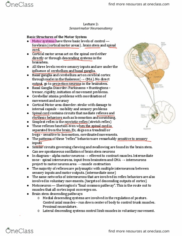
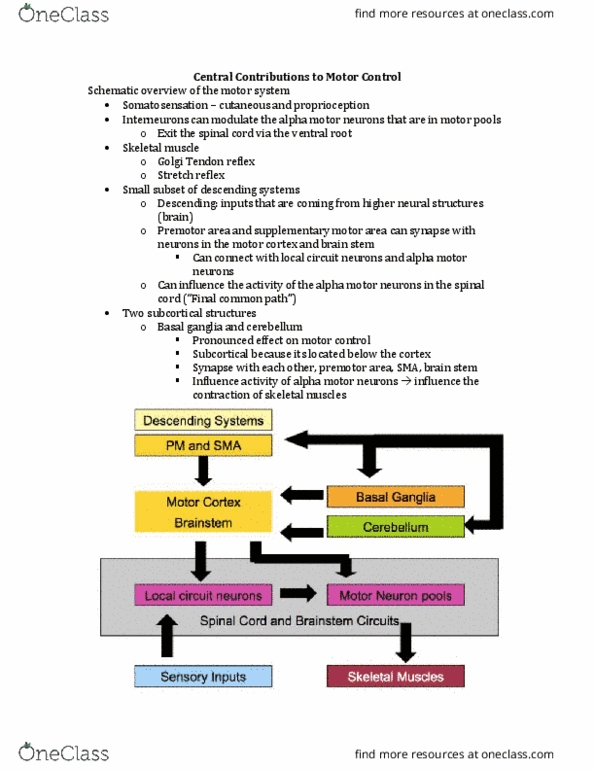
Control of movement:
- An important concept of neuronal pathways: set of
neurons connected in serial fashion. There are
ascending and descending pathways
- In descending pathways:
o The structures (set of neurons) responsible for
motor control (called motor centres) include
nuclei in the brainstem (low level), basal ganglia
and motor cortex (medium level), and
association areas of neocortex (high level).
o The motor centres (= upper motor neurons)
control activity of lower motor neurons in the
spinal cord
o The motor centres perform appropriately if
receiving continuous input from sensory system.
sensory-motor system participates in movement control
Major steps in neuro-muscular transmission:
- Spinal motor neurons have cell bodies in ventral horn of spinal
cord. Their axons leave spinal cord to reach appropriate muscle
fibres and form neuro-usula sapses ed plates
- AP of excited motor neurons are conducted to motor nerve
endings, ACh released
- ACh produces a loalised ed plate potetial siila to euo
EPSP) in muscle
- Single end plate potential usually triggers a muscle AP, which
spreads over entire muscle and triggers muscle contraction
Motor units and motor neuron pool:
- A single muscle, e.g. biceps, is composed of thousands of skeletal muscle fibres
- Each muscle fibre is innervated by a single motor neuron. However, 1 motor neuron
innervates many fibres (= motor unit).
o In extensors of the leg – 1 motor neuron innervate 1000 muscle fibres
o In muscles controlling finger/eye movement: only few muscle fibres
- Musles ith lage ue of sall oto uits: fiel otolled uppe oto
euos i ai ia loe oto euos i spial od
- Collection of lower motor neurons innervating muscle fibres of a muscle is called the
oto euo pool of that usle
find more resources at oneclass.com
find more resources at oneclass.com

Synergists and antagonists:
- Flexion bends limb ( joint angle)
- Extension straightens limb ( joint angle)
- Movements usually by groups of muscles working together (synergists)
- Antagonists: muscles which perform opposite actions
Proprioceptors: Muscle spindle (sensory receptor) →
- Group of small modified muscle fibres (8-10 intrafusal muscle fibres)
- Senosry nerves of group Ia (& group II) afferents innervate the
intrafusal fibres
- Every muscle contains dozens of muscle spindles, in parallel with
extrafusal muscle fibres (which signal muscle length)
Stretch reflex: example of monosynaptic connection, & reciprocal innervation
- Stretching a muscle spindle of flexor muscle (biceps) → firing of Ia
sensory neuron & firing of motor neurons that innervate the same
muscle (no interneurons monosynaptic connection)
- Ia afferents excite motor neurons innervating synergistic muscle, and
indirectly (via inhibitory interneurons) inhibit motor neurons innervating
antagonistic muscle reciprocal innervation (simultaneous relaxation of antagonist)
- Negative feedback loop used to maintain muscle length at desired value
- Skeletal muscles are always under some degree of stretch, stretch reflex circuits are
responsible for steady level of tension in muscle (muscle tone)
- Stretch reflexes do not involve brain, initiates muscle contraction within 25ms
motor neurons modulate the level of excitability of muscle spindle
- When flexor contracts, muscle spidle is uloaded Ia affeets stop fiig, see A )
-
- However, activation of motor neurons, which terminate on contractile poles of
intrafusal fibres, evokes their contraction, thus maintaining tension on middle region
where sensory axons terminates (see B )
- Co-activation of and motor neurons allows spindles to function at all length
during movement
find more resources at oneclass.com
find more resources at oneclass.com
Document Summary
Learning objectives: spinal motor neurons, proprioceptors: muscle spindles & golgi tendon organs, neural circuits of spinal reflexes: stretch & withdrawal reflexes, role of major regions of the brain (brainstem, cerebellum, basal ganglia, and cortex) in control of movement. An important concept of neuronal pathways: set of neurons connected in serial fashion. Spinal motor neurons have cell bodies in ventral horn of spinal cord. Their axons leave spinal cord to reach appropriate muscle fibres and form neuro-(cid:373)us(cid:272)ula(cid:396) s(cid:455)(cid:374)apses (cid:894)(cid:858)e(cid:374)d plates(cid:859)(cid:895) Ap of excited motor neurons are conducted to motor nerve endings, ach released. Ach produces a lo(cid:272)alised (cid:858)e(cid:374)d plate pote(cid:374)tial(cid:859) (cid:894)si(cid:373)ila(cid:396) to (cid:374)eu(cid:396)o(cid:374) Single end plate potential usually triggers a muscle ap, which spreads over entire muscle and triggers muscle contraction. A single muscle, e. g. biceps, is composed of thousands of skeletal muscle fibres. Each muscle fibre is innervated by a single motor neuron. However, 1 motor neuron innervates many fibres (= motor unit).