1001NSC Lecture Notes - Lecture 12: Carbonic Anhydrase, Trachea, Gas Exchange

20 Jun 2018
School
Department
Course
Professor

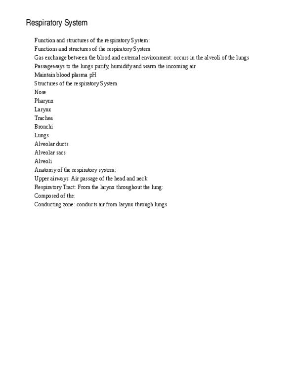
The Respiratory System
Consists of: nasal cavity, oral cavity, pharynx, larynx, trachea, lungs, bronchial tree, alveoli,
pleurae (casing of each lung)
Lungs:
Right side: 3 lobes
Left side: 2 lobes
Entry and exit points of blood vessels are situated in hilus of each lung
Pleural membranes and Plural Cavity
Visceral Pleura covers the lungs
Parietal Pleura lines the ribcage and covers the upper surface of diaphragm
Pleural cavity is the potential space between the ribs and lungs
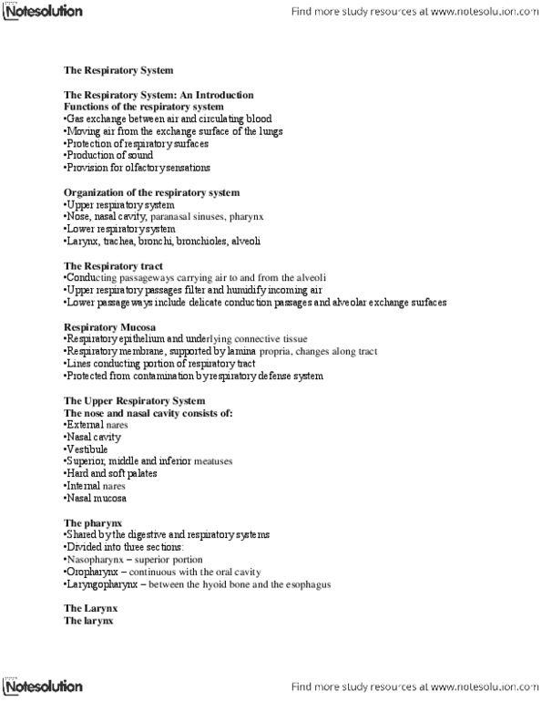
Respiratory Regions
Conducting Region (the bronchial region)
Comprises: nose, nasal cavity, oral cavity, pharynx, larynx, trachea, primary bronchi,
secondary and tertiary bronchi, bronchioles, terminal bronchioles
Lining of conducting region:
Blood vessels warm the air entering the lungs
Mucus (secreted by goblet cells): traps dust, microbes and foreign particles, moistens the
air (stops respiratory system from drying out)
Cilia: columnar cells in 300 cilia/cell, 14 cycles, proper mucus, sweep in one direction
propelling mucus and debris. Lots of cilia activity results in respiratory infections and
pneumonia
Cartilage: hyaline cartilage within alls of trachea and bronchi prevents collapse of the air
passages
Smooth muscle: increasing amounts until complete player present in bronchioles. Allows
regulation of airflow by bronchioconstriction/ bronchiodilation
Pharynx:
Nasopharynx
Oropharynx
Laryngopharnx
Uvula: prevents food from going up into the back of nose
Epiglottis: prevents food from entering trachea and lungs
Respiratory region:
Site of gas exchange between atmosphere and body
Respiratory bronchioles
Alveolar ducts
Alveolar sacs (clusters of alveoli)
Alveoli (site of gas exchange)
Respiratory membrane:
Composed of
Type I alveolar cell (alveolar epithelial cell)
Blood capillary endothelial cell (wall of capillary)
Small amounts of collagen
Breathing:
Movement of air into and out of the lungs
Under voluntary and involuntary control
Occurs in response to pressure changes
find more resources at oneclass.com
find more resources at oneclass.com
Document Summary
Consists of: nasal cavity, oral cavity, pharynx, larynx, trachea, lungs, bronchial tree, alveoli, pleurae (casing of each lung) Entry and exit points of blood vessels are situated in hilus of each lung. Parietal pleura lines the ribcage and covers the upper surface of diaphragm. Pleural cavity is the potential space between the ribs and lungs. Comprises: nose, nasal cavity, oral cavity, pharynx, larynx, trachea, primary bronchi, secondary and tertiary bronchi, bronchioles, terminal bronchioles. Blood vessels warm the air entering the lungs. Mucus (secreted by goblet cells): traps dust, microbes and foreign particles, moistens the air (stops respiratory system from drying out) Cilia: columnar cells in 300 cilia/cell, 14 cycles, proper mucus, sweep in one direction propelling mucus and debris. Lots of cilia activity results in respiratory infections and pneumonia. Cartilage: hyaline cartilage within alls of trachea and bronchi prevents collapse of the air passages. Smooth muscle: increasing amounts until complete player present in bronchioles.