PS 1001:03 Lecture Notes - Lecture 13: Condyloid Process, Mandibular Fossa, Articular Disk


Articulation between mandible and temporal bone
-
Mandible = u shaped jaw bone
-
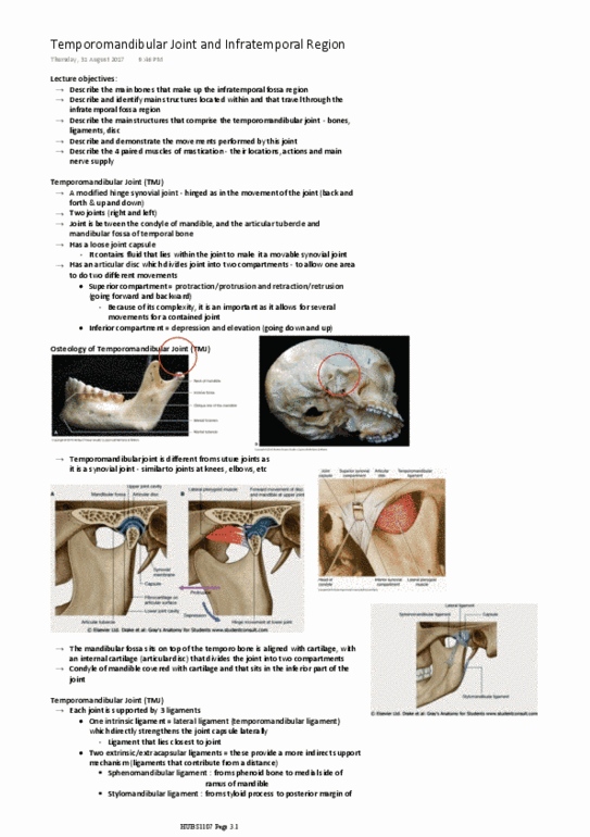
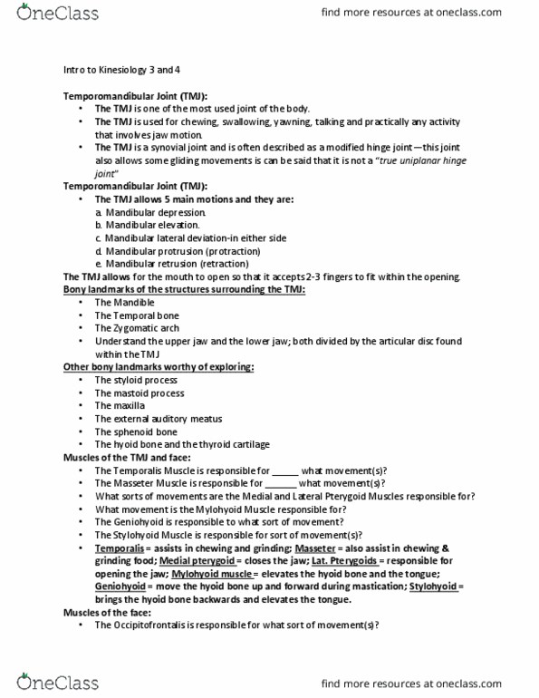
Temporomandibular joint
Condyle of mandible with mandibular fossa of temporal bone
-
Contains interarticular disc enclosed by a joint capsule
-
Functional anatomy
Screen clipping taken: 2/04/2018 10:03 PM
Potential source acute chronic pain in head and neck, which can be attributed to joint and
muscles surrounding it
-
Headaches
○
Altered taste
○
Tinnitus
○
Dysfunctions of joint include
-
Mastication - chewing
○
Speech
○
Participates in essential functions
-
Provides cephalic articular surface for TMJ
○
Temporal bone
-
Easily palpated inferior and posterior to the external auditory meatus
○
Close proximity of inner, middle and outer ear to TMJ may explain why some people
with TMJ dysfunction can impaired hearing
○
May complain of ear pain, tinnitus or impaired hearing
○
Mastoid process
-
Marks junction of the body and the ramus
○
Can be easily palpated the posterior aspect of the jaw on either side of the face
○
Angles of mandible important landmarks - because lying superior, the posterior tips of
the hyoid bone and inferior and anterior to the transverse processes to the atlas
○
So articular surfaces covered by articular cartilage
▪
But articular cartilage consists of fibro cartilage rather than hyaline cartilage,
which is relatively avascular which has to be nourished and lubricated by synovial
fluid
▪
Presence of fibro cartilage suggests the TMJ has to sustain large forces e.g. when
chewing or biting into food
▪
Large forces sustained mostly in chewing actions
▪
Condyles shaped like footballs cut in half, that tilt anteriorly and medially towards each
other
○
Mandible
-
L3 - TMJ
Monday, 2 April 2018
10:02 PM
week 6 Page 1

Mandibular fossa of temporal bone - articular surface is formed by the sloped anterior wall of
the fossa
-
Condyles of the mandible oriented anteriorly and medially
-
Articular surfaces
Screen clipping taken: 2/04/2018 10:13 PM
Screen clipping taken: 2/04/2018 10:13 PM
3 dimensional motion
-
Ie. Opening and closing, sagittal plane
▪
Medial-lateral (A)
○
Rotation about
-
Motion at TMJ
week 6 Page 2
Document Summary
Condyle of mandible with mandibular fossa of temporal bone. Contains interarticular disc enclosed by a joint capsule. Potential source acute chronic pain in head and neck, which can be attributed to joint and muscles surrounding it. Easily palpated inferior and posterior to the external auditory meatus. Close proximity of inner, middle and outer ear to tmj may explain why some people with tmj dysfunction can impaired hearing. May complain of ear pain, tinnitus or impaired hearing. Marks junction of the body and the ramus. Can be easily palpated the posterior aspect of the jaw on either side of the face. Angles of mandible important landmarks - because lying superior, the posterior tips of the hyoid bone and inferior and anterior to the transverse processes to the atlas. Condyles shaped like footballs cut in half, that tilt anteriorly and medially towards each other.