PSYC10003 Lecture Notes - Lecture 8: Incus, Eardrum, Basilar Membrane

14 Jun 2018
School
Department
Course
Professor

MBB1 – Lecture 8
Auditory and somatosensory systems
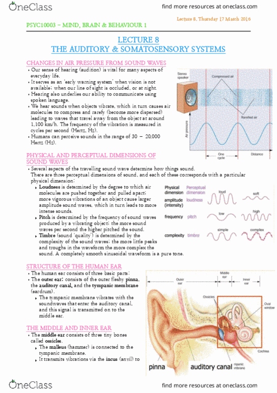
Changes in air pressure from sound waves
• We hear sounds when objects vibrate, which caused air
molecules to compress and rarefy (become more dispersed)
• Frequency of the vibration is measured in cycles per second
(Hertz, Hz)
Physical and perceptual dimensions of sound waves
3 perceptual dimensions of sound that correspond to a physical
dimension:
1. loudness – determined by degree to which air molecules or
pushed together or apart → more vigorous vibrations of object causes larger
amplitude waves, leads to more intense sound
2. pitch - determined by frequency of sound waves → more waves per second, higher
pitch
3. timbre – quality is determined by complexity of waves → more little peaks/troughs,
the more complex
• Higher – more compressed
• Lower – more rarified
• We can change the rate the compressed phase gets to us
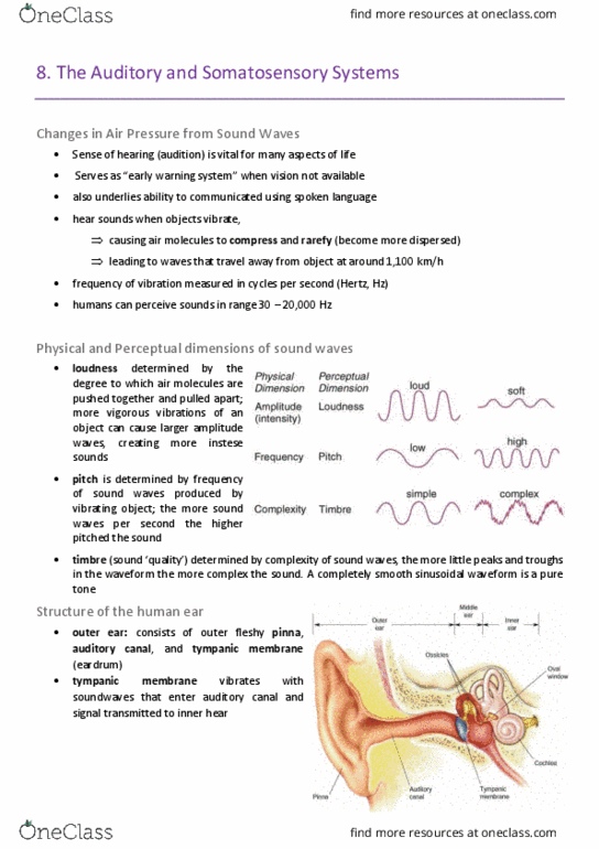
Structure of human ear
• outer ear:
o Pinna – helps focus sound waves from the source
o Auditory canal
o Tympanic membrane (eardrum) – vibrates with soundwaves entering
auditory canal - movement of eardrum causes movement of ossicles
• Middle ear:
o Ossicles – three tiny bones
▪ Malleus (hammer) is connected to tympanic membrane and transmits
vibrations via the incus (anvil) to the stapes (stirrup), which is
connected to the cochlea
• Inner ear:
o Cochlea – contains receptors for analysing sounds
▪ Bony structure with 2 small membranes
forming windows on its fluid-filled interior
▪ Stapes connected to oval window
▪ Sound waves cause stapes to move, & move
fluid over receptors in cochlea
▪ Another membrane needed to allow fluid to
move – round window
• Basilar membrane within cochlea – sheet of tissue
containing the auditory receptors
o sits in centre of cochlea, runs from its base to its
apex
find more resources at oneclass.com
find more resources at oneclass.com

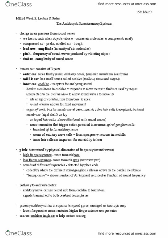
Organ of Corti
• cross-section across longitudinal axis of cochlea reveals three inner chambers, each
of which is filled with fluid
• on floor of centre chamber is the organ of Corti, which runs the length of the cochlea
• composed of basilar membrane (BM) at its base, receptors in the middle called hair
cells (inner and outer), and a rigid shelf over the top called the tectorial membrane
hair cells and stereocilia
• on top of the hair cells are tiny filaments called stereocilia
• sound waves cause BM to move relative to tectorial membrane above it
o this motion effectively bends the stereocilia, either by direct contact with
tectorial membrane (for outer hair cells), or by fluid motion induced by
movement of BM
o bending of stereocilia of hair cells is what produces receptor potentials that
convert sounds waves into neural signals
scanning micrograph image of organ of Corti where tectorial membrane has been cut away
to see hair cells
find more resources at oneclass.com
find more resources at oneclass.com