PSYC20006 Lecture Notes - Lecture 7: Hemoglobin, Seiji Ogawa, Superposition Principle

2 Jul 2018
School
Department
Course
Professor

PSYC20006 Biological Psychology
1
WEEKS 1 - 5: STATISTICS & IMAGING METHODS
LECTURE 7 – 8 (W4):
Functional Magnetic Resonance Imaging (fMRI) Methods
Why fMRI is used in psychology
• Want to know things about cognitive processes
o Cognition happens in the brain
o Use reverse inference from the fMRI → draw conclusions about cognitive
processes from presence of activation
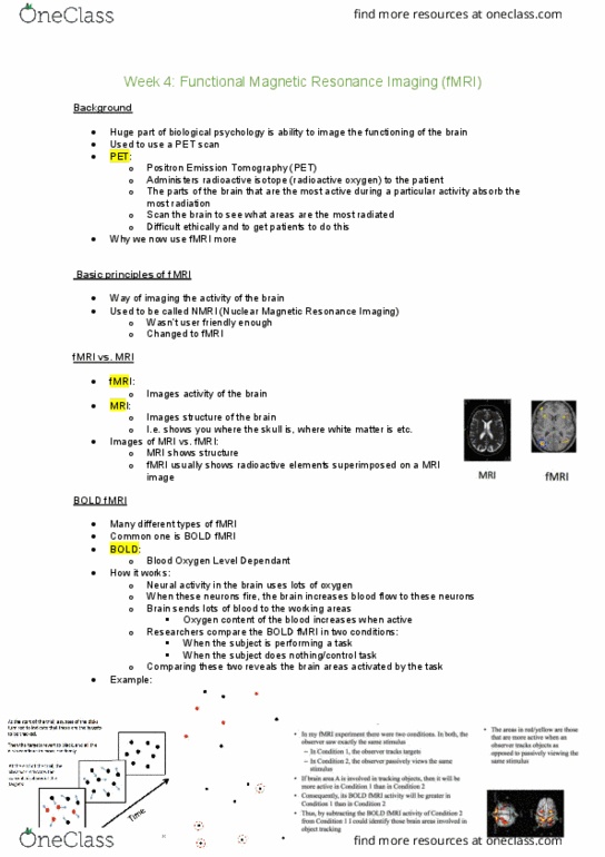
• 1st method was Positron Emission Tomography (PET)
o Administer radioactive isotope to patient (e.g. oxygen 15)
▪ Exposes patient to significant amount of ionising radiation
• fMRI: originally NMRI (nuclear MRI)
o more commonly used now; no radiation
fMRI
• 1.5 - 9 Tesla mag field; usually 3T for imaging
o VERY STRONG FIELD (Earth’s magnetic field = 65 micro T)
o Don’t take in metal
• Participant placed on bed and moved into magnet
• Experiment can be controlled from outside scanner room
• Head coil
o Participants can see projection (usually computer-controlled experiment)
via mirrors mounted on head coil
o Give responses via scanner-compatible keys, joystick, touchpad etc.
o Used to send radio frequency pulses & functions as a receiver
o Fixes head to avoid movement
MRI: Basic physics
• Images the structure of the brain
• Over 70% of brain consists of water
o H atoms (H+ protons) can be thought of as small bar magnets
▪ Precessing like a spinning top around an axis
• Protons’ random spin directions aligned parallel or anti-parallel to externally
applied very strong magnetic field in MRI scanner
o Not all perfectly aligned or static
o Precessing in a random fashion
o Precessing frequency (resonance frequency) depends on strength of
magnetic field
• Z-axis: axis the magnetisation is built up in the scanner
o Magnetisation here can’t be measured
o Need to tilt magnetisation vector
PSYC20006 Biological Psychology
2
▪ Radiofrequency (RF) pulse applied perpendicular to mag field
• Amplitude matches proton precession frequency
• Causes protons to absorb energy
• Tilts magnetic vector to transversal plane
• Aligns precession of spins so protons’ rotations in phase
• Transversely rotating magnetising vector recorded as signal
o Now switch off RF pulse
• Relaxation: transversal magnetisation disappears, longitudinal magnetisation re-
established
o Summed effect of many protons doing this measured in this phase
• Transversal magnetisation decays at different speeds depending on tissue
o Because of differences in density of protons
▪ Lose coherence - influenced by other protons in environment
o Signals from different protons will get out of phase with each other & begin
to cancel each other out
o Structural brain image depends on when signal recorded in process
MRI: Mechanisms of scanning
• Reconstructing brain images
o Need to decide exactly where the signal comes from
▪ Cannot excite entire brain with RF pulses at once because couldn’t
reconstruct source of measured signal
o Knowing protons absorb energy from RF pulses only when frequency of RF
pulses match proton’s precession frequency
o Procedure
▪ Use gradients to cause magnetic field to vary linearly to cause
resonance frequency throughout brain
• RF pulse of specific frequency will only cite one slice of the
brain (where the resonance frequency of the protons matches
the frequency of the RF pulse)
▪ Slice selecting gradient
• Divide brain into ‘slices’
• To be able to vary gradient field along z-axis and know
different slices exposed to different field strengths
▪ Frequency encoding gradient
• 2nd gradient, to change magnetic field within slice
• During readout, vary gradient along y-axis
• Means protons in each slice have different precession
frequencies
• Gives y-coordinate of measured signal
▪ Phase encoding gradient
• Briefly using gradient along x-axis causes protons to ‘speed
up’ precession
• According to strength of magnetic field for short time