BMS1052 Lecture Notes - Lecture 23: Developmental Neurobiology, Neural Crest, Neural Plate

5 May 2018
School
Department
Course
Professor

Lecture 23 Developmental neurobiology 1,2 -Sonja Mckeown
Lecture objectives
• At the end of these lectures you will be able to:
• Explain why understanding development is important in appreciating nervous
system anatomy and function
• Identify the developmental origin of the nervous system.
• Describe the stages of development of the central nervous system, namely neural
induction, neurulation, neural patterning, neurogenesis, neuron migration, axon
guidance, synaptogenesis, neuronal death and gliogenesis.
• Describe the origins of the peripheral nervous system, including migration of the
neural crest and major neural derivatives.
• Identify some of the events that can go wrong during development and
perturbations of development that can occur.
• Explain the concept of critical periods in brain development.
Why study development?
• Understand the basis of anatomy, and function.
• Understand how things can go wrong – perturbations of development.
• Understand how things can go wrong – adult disease (eg cancer).
• Understand the basis behind seemingly diverse phenotypes.
• Stem cells.
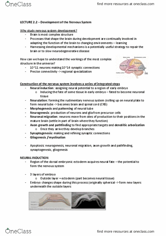
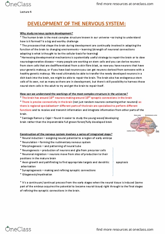
Early development- gastrulation
The developing embryo undergoes gastrulation. This creates three layered embryo
(ectoderm, mesoderm, endoderm). Ectoderm gives rise to neural tissue. This is the setting
up for neural induction.
Neural induction the
neural plate develops from
the top ectoderm, the top
layer of the embryo. The
nervous system develops
from the ectoderm. A
specialised region froms the
neural plate
find more resources at oneclass.com
find more resources at oneclass.com

The ectoderm, the top layer of the embryo, gives rise to different tissues in the adult body.
The most obvious is skin. But also, it produces neural tissue – from several different
populations of ectoderm. We focus mostly on the neural plate, which forms the brain and
spinal cord, but the major source of the peripheral nervous system, which includes sensory
neurons and peripheral autonomic neurons, is the neural crest. Neural crest also form
other important structures, including bones and cartilage of the face and jaw, and
melanocytes in the skin. There are also several placodes of the ectoderm, which develop a
bit like the neural plate only much smaller, and form some peripheral sensory neurons in
the head region. (also other placodes which form the lens, olfactory epithelium (primary
olfactory neurons), and inner ear – otic placode).
Neural induction and neural crest induction
• Signals converge on the middle region of the ectoderm and induce it to become
neural tissue. The signals come from the ectoderm, the node (a signalling centre),
and the underlying mesoderm. These signals act on the middle of the ectoderm and
cause it to become the neural plate.
• The border region between the neural plate and ectoderm is induced to become
neural crest. Induction means that the gene expression patterns of the tissue
changes – in particular the tissues express particular transcription factors, which
regulate expression of other genes. This makes the tissue different from
surrounding regions, and capable of differentiating into (becoming) a specific
structure or tissue.
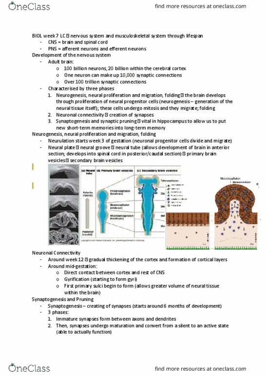
Neurulation when flat plate folds into a tube (formation of neural tube)
• The neural plate rolls up into a tube.
• The centre (lumen) of the tube is filled with fluid. The fluid in the lumen of the neural
tube is cerebrospinal fluid (CSF), which is found within the brain vesicles (created by
the choroid plexus) and in the subarachnoid space in the meninges.
find more resources at oneclass.com
find more resources at oneclass.com

Neural tube defects
• Craniorachischisis : neural tube completely open. Where the neural tube is exposed to
amniotic fluid, the neuroepithelium degenerates.
• Anencephaly: failure of rostal neuropore closure
• Spina bifida : failure of caudal neuropore closure damage to the spinal cord, and lack of
muscle control in the lower part of the body.
• Spinal dysraphism (closed spina bifada) : bony covering of caudal vertebrae incomplete
Neural tube patterning
• Parts of the neural tube swell to become vesicles.
• These vesicles form different parts of the brain (anterior- posterior patterning).
How is one end of the embryo made to be different to the other end? The neural tube
has to be patterned so there is a brain at the head end and spinal cord along the trunk.
Different parts of the brain also need to be patterned. Patterning occurs in the anterior-
posterior (top to bottom, rostral to caudal), and dorsal-ventral (back-front) axes.
• The neural tube joins up first in the
middle, and zippers up to closure
points.
• Neural tube defects occur when the
neural tube fails to close.
There are specific sites of the neural tube
where it joins up to form a tube (closure
points), and then progresses toward the
remaining open neuropores (rostral/anterior
and caudal/posterior). The main closure point
is at the hindbrain. There are different types
of neural tube defects, depending on where
the neural tube fails to close. The most
common is spina bifida, where the neural
tube has failed to close at the caudal
neuropore.
find more resources at oneclass.com
find more resources at oneclass.com
Document Summary
Lecture objectives: at the end of these lectures you will be able to, explain why understanding development is important in appreciating nervous system anatomy and function. Identify some of the events that can go wrong during development and perturbations of development that can occur: explain the concept of critical periods in brain development. This creates three layered embryo (ectoderm, mesoderm, endoderm). This is the setting up for neural induction. Neural induction the neural plate develops from the top ectoderm, the top layer of the embryo. The ectoderm, the top layer of the embryo, gives rise to different tissues in the adult body. But also, it produces neural tissue from several different populations of ectoderm. We focus mostly on the neural plate, which forms the brain and spinal cord, but the major source of the peripheral nervous system, which includes sensory neurons and peripheral autonomic neurons, is the neural crest.