BMS1052 Lecture 17: Lecture 17,18,19 Control of movement 2 → controlling contractions

5 May 2018
School
Department
Course
Professor

Lecture 17 Control of movement 2 controlling contractions
At the end of this lecture you should be able to:
- Explain the molecular mechanisms underlying force summation and excitation-
contraction coupling
- use the sliding filament theory to explain the length-tension relationship in
sarcomeres
- define isotonic, isometric, concentric and eccentric contractions
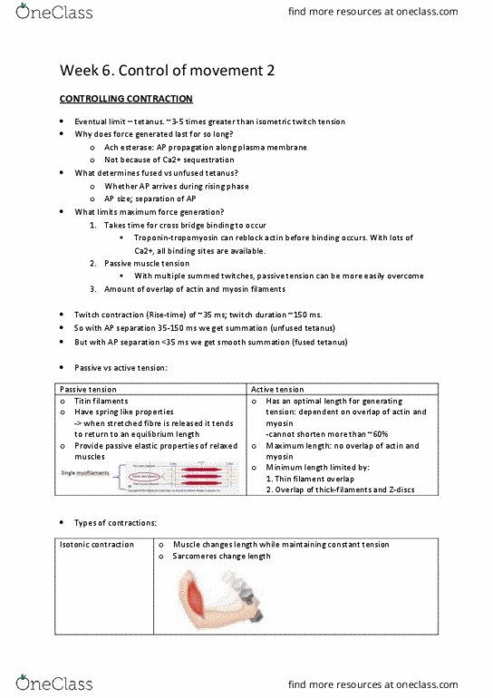
- list the properties of the three types of skeletal muscle fibers
- relate the function of different fiber types to their underlying properties
- explain the results of crossed-innervation experiments
Excitation- Contraction coupling
• Excitation stimulation by a neuron
• Contraction interaction b/w actin and myosin
Muscle is attached to bone via a tendon.
Fascicle - Bundle of fibres surrounded by connective tissue.
1. Blood vessels and nerves are integrated with this connective tissue
2. Connective tissue is tough – helps resist passive muscle stretch and prevent muscle
tearing
3. Helps distribute force from muscle fibres to tendon and bone.
Whole muscle = made up of multiple muscle fibres
Each muscle fiber is made up of multiple contracticle subunits called myofibrils
Fro UP’s leture otes:
Functions of the connective tissue covering are:
1. The perimysium provides a conduit for blood vessels and nerves supplying
the muscle fibres.
2. The perimysium resists passive stretching of the muscle and ensures forces
are distributed uniformly to prevent tearing of muscle fibres.
3. The endomysium through its lateral connections to adjacent muscle fibres
conveys part of the contractile force to the tendon.
A myofibril is 1-2 um in diameter.
Sarcomere = basic functional unit of a muscle – repeating section of actin and myosin
filaments. These account for striated appearance. Ends at z lines
Each sarcomere is ~1.5-4 um long,
Synapse = the connection b/w the neuron and the effector cell (in this case the muscle)
Synaptic cleft = the space in b/w (the gap where the NT is released)
find more resources at oneclass.com
find more resources at oneclass.com

Critical steps:
1 - Motor neuron action potential
3 – Acetylcholine (Ach) release allows influx of small cations (notably Na+) (K+ will leak out)
cell becomes depolarised and all the positives become negatives and vice versa.
5 – Na+ entry in motor end plate triggers opening of adjacent voltage gated Na+ channels
8 – Muscle action potential propagates along plasma membrane, which is electrically active.
Depedig o the tetook soeties ou’ll read ACH-receptors as Nicotinic receptors
Note that to prevent ongoing AP and contraction, acetylcholine-esterase breaks down Ach.
An aside: atropine, is a competitive antagonist of the muscarinic Ach receptors. Historically,
atropine derived from deadly nightshade was applied directly to the eyes of women to
make them more beautiful. Who can think what it might be doing? Leads to pupil dilation,
due to relaxation of iris.
Excitation- contraction coupling
Dihydropyridine receptor is a voltage-gated Ca2+ channel – it opens in response to AP
DHP receptor and ryanodine receptor are physically coupled Ca2+ channels
AP increases cytosolic calcium levels because Ca2+ is release from the SR
Ca2+-ATPases pump Ca2+ ions from cytosol back into the SR
SO the depolarisation will spread to the T-tubules and within the t-tubles are receptors
know as the DHP Dihydropyridine receptor is voltage regulated, what does that mean?
It means it changes shape when the voltage changes.
That eas that he the AP oes, there’s a release of ACH influx of NA2+ then
there’s a spread of atio potetial to the t-tubules
When the DHP receptors change shape they connect to the Ryanodine receptor and open it
like a treasure box and then inside it spills out Ca2+ from the SR
Remember – cytosolic, or intracellular Ca2+ concentration is normally kept very low.
What ions are DHP and ryanodine receptors permeable to? Ca2+
Action potential propagates along surface of plasma membrane (grey) and down into
transverse tubules.
find more resources at oneclass.com
find more resources at oneclass.com

Summary
1/ Action potential travels down the motor neuron
2/ ACH is released in the NMJ
3/ ACH binds to the ACH receptors
4/ Influx of sodium
5/ Wave of depolarization
6/ AP goes to t-tubles
7/ Voltage gated DHP receptors open/ change in shape
8/ Opens the Ryanodine receptor
9/ SR releases Ca2+
Crossbridge cycling
i) Myosin head is initially uoud, ut eergised
ii) Actin binding site is blocked by tropomyosin
iii) Ca2+ binds to troponin, moving tropomyosin, unlocking cross bridge binding sites
iv) Myosin cross-bridges bind to actin
v) power stroke: myosin head rotates release ADP and P
vi) ATP binding to myosin breaks A-M linkage
vii) Bound ATP is split, re-energising myosin head.
Initially, unbound, but cross-ridge is eergised, iplig that it ould easil rotate,
leading to force generation or contraction.
Myosin is unable to bind to actin filament because it is blocked by tropomyosin.
Myosin head at this particular state is also energised ATP is bound hydrolysed to ADP
ad P COCKED positio
Sarcomere- sliding filament theory
The sarcomere is the individual contractile subunit, when you look at the ofiril there’s
heaps of little sarcomeres.
When every single sarcomere contracts the myofibril will contract and shorten the
muscle will then contract and shorten this is what produces a change in length for
muscles.
The ROLE OF ATP
1. Hydrolysis of ATP osi oked positio
2. ATP binding to myosin dissociates cross-bridges bound to actin
3. Hydrolysis of ATP by the Ca2+-ATPase actively transports calcium ions in the
sarcoplasmic reticulum, allowing relaxation
find more resources at oneclass.com
find more resources at oneclass.com
Document Summary
Lecture 17 control of movement 2 controlling contractions. Excitation- contraction coupling: excitation stimulation by a neuron, contraction interaction b/w actin and myosin. Muscle is attached to bone via a tendon. Whole muscle = made up of multiple muscle fibres. Each muscle fiber is made up of multiple contracticle subunits called myofibrils. Sarcomere = basic functional unit of a muscle repeating section of actin and myosin filaments. Synapse = the connection b/w the neuron and the effector cell (in this case the muscle) Synaptic cleft = the space in b/w (the gap where the nt is released) 3 acetylcholine (ach) release allows influx of small cations (notably na+) (k+ will leak out) Cell becomes depolarised and all the positives become negatives and vice versa. 5 na+ entry in motor end plate triggers opening of adjacent voltage gated na+ channels. 8 muscle action potential propagates along plasma membrane, which is electrically active.