BMS1052 Lecture Notes - Lecture 13: Lateral Geniculate Nucleus, Pupillary Light Reflex, Suprachiasmatic Nucleus

5 May 2018
School
Department
Course
Professor

Lecture 13 WEEK 5 Vision and the eye 2
Learning objectives
1. Describe the structure and benefits of center-surround receptive fields
2. Describe the properties of magno-, parvo- & konio-cellular retino-thalamic
projections
3. Describe how the visual field is assembled from inputs from the two eyes, and the
effects of lesions in different positions along the visual pathway
4. Describe the functional organisation in V1 and the basic properties of simple and
complex cells
5. Identify the major functional roles of the dorsal and ventral visual streams
Lecture outline
1. Center-surround receptive fields
2. Where does information from the retina go? The retino-thalamic-cortical pathway
3. The thalamic lateral geniculate nucleus
4. Primary visual cortex
5. The two visual streams
Receptive fields and bipolar cells
A reeptie field is the regio of spae i hih light hages affet a ell’s erae
potential.
Bipolar cells have an antagonistic, center-surround receptive field structure.
OFF bipolar cells
- hyperpolarised by light in their RF center
- Preserve sign of photoreceptors
- Depolarised by light in their RF surround
Remember, in the dark, photoreceptors are
depolarised and tonically active, and increased light hyperpolarises them (reduces their
output).
i.e. Light off = more glutamate; light on = less glutamate.
There are two types of bipolar cells
OFF cell – Sign-preserving. Glutamate from photoreceptors is excitatory (depolarises cells) -
bipolar cells preserve photoreceptor signal => Increased illumination = less glutamate =>
hyperpolarises cell.
ON cell – Sign inverting - Glutamate from photoreceptors is inhibitory (hyperpolarises) –
bipolar cells invert photoreceptor signal => increased illumination = less glutamate =>
depolarises cell. ON cell
What iruit aounts for a ipolar ell’s enter-surround RF?
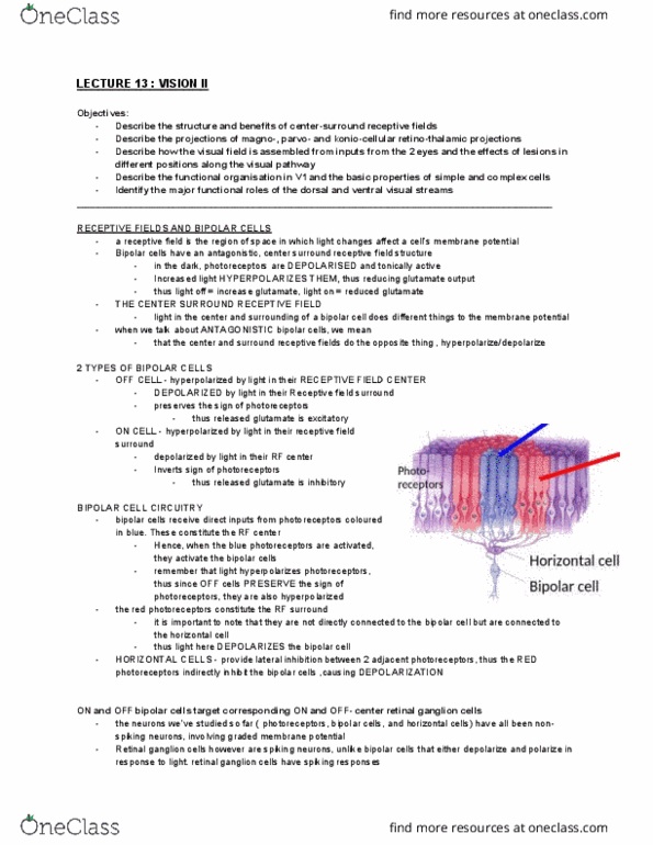
Photoreeptors otriutig to ipolar ell’s RF center
These are directly connected to the bipolar cell.
Light here hyperpolarises an OFF bipolar cell
ON bipolar cells
- depolarised by light in their RF
center
- Invert sign of photoreceptors
- Hyperpolarised by light in their RF
surround
find more resources at oneclass.com
find more resources at oneclass.com

Photoreeptors otriutig to ipolar ell’s RF surround.
These are only connected to the horizontal cell
Light here depolarises an OFF bipolar cell
(Basic) summary of cell types in retina
Photoreceptors (rods/cones)
- light transduction via G-protein coupled opsin molecule
- depolarised in darkness
- non-spiking; glutamate release decreases as light levels increase
- Three cone classes have different wavelength sensitivity
- no rods in fovea; fewer cones in periphery
Bipolar cells
• OFF-bipolar cells are hyperpolarised by light on their RF center
• ON-bipolar cells are depolarised by light on their RF center
• center-surround receptive field
• graded membrane potentials (non-spiking)
Horizontal cells
- bidirectional, inhibitory connections to photoreceptors
- mediate bipolar cell receptive field surrounds
-
Retinal ganglion cells
- center-surround receptive fields
- spiking responses
- retinal output cells
- convergence ratio of inputs from photoreceptors affects visual acuity
Center-surround receptive fields can (mostly)
explain the illusions
Center-surround organisation (lateral inhibition) is:
- one of the fundamental mechanisms in all sensory systems
- improves spatial localisation
- improves stimulus identification
- Stimulus borders, edges and changes are useful and interesting
- not constant stimulation.
Where does information from the retina go? The retino-thalamic-cortical pathway
Functional segregation:
retinal ganglion cells project to many places
Lateral geniculate nucleus – (LGN) gateway to cortex and conscious vision
Pretectum – reflexive eye movements and pupil size.
Superior colliculus - controls eye and head orienting responses
Suprachiasmatic nucleus in hypothalamus – circadian rhythms
Also, the Pulvinar, pregeniculate nucleus and accessory optic system
find more resources at oneclass.com
find more resources at oneclass.com

Since ~90% of retinal projection is to the LGN, which subsequently sends significant
projections to the cortex, we will focus on the retino-thalamo-cortical pathway for the rest
of the lecture.
Be sure to understand the difference between what is seen by the left / right eyes, and
visual hemifield – left or right of the vertical meridian.
For the sole purpose of confusing students and providing lecturers something to put in
exams, there is a partial decussation of the visual fibers.
So the left visual field is processed by the right side of the brain.
Put another way:
- The image on the right (nasal) portion of the left eye projects to the right side of the
brain
- the image on the right (temporal) portion of the right eye, projects to the right side
of the brain
Fibres from each nasal hemiretina
decussate in the optic chiasm.
Thus:
- The left visual cortex represents the right visual field.
- The right visual cortex represents the left visual field.
Partial decussation – each eye receives information both sides of the visual world.
However, each side of the brain only receives information from one side of the visual
world.
Distinguish nasal and temporal retina – vertical meridian is a vertical line through your
point of focus.
“o if I’ lookig straight ahead, eerthig o the right, is proessed the left side of
my retina = left temporal; right nasal.
Different ganglion cell types tile the retina
- functional segregation
• >12 parallel circuits, with unique classes of ganglion cell
• Each circuit receives inputs from the same cone photoreceptors,
but the inputs are processed in different ways.
Retinal outputs demonstrate functional and anatomical segregation
Midget cells (P-type cells)
- 80-90% of ganglion cells
- small cell bodies, dendritic arbors, receptive fields
- sensitive to fine stimulus features
Left
Right
Visual
Fields
find more resources at oneclass.com
find more resources at oneclass.com
Document Summary
Lecture 13 week 5 vision and the eye 2. The retino-thalamic-cortical pathway: the thalamic lateral geniculate nucleus, primary visual cortex, the two visual streams. A re(cid:272)epti(cid:448)e field is the regio(cid:374) of spa(cid:272)e i(cid:374) (cid:449)hi(cid:272)h light (cid:272)ha(cid:374)ges affe(cid:272)t a (cid:272)ell"s (cid:373)e(cid:373)(cid:271)ra(cid:374)e potential. Bipolar cells have an antagonistic, center-surround receptive field structure. Hyperpolarised by light in their rf center. Depolarised by light in their rf surround. Depolarised by light in their rf center. Hyperpolarised by light in their rf surround. Remember, in the dark, photoreceptors are depolarised and tonically active, and increased light hyperpolarises them (reduces their output). i. e. light off = more glutamate; light on = less glutamate. Glutamate from photoreceptors is excitatory (depolarises cells) - bipolar cells preserve photoreceptor signal => increased illumination = less glutamate => hyperpolarises cell. On cell sign inverting - glutamate from photoreceptors is inhibitory (hyperpolarises) bipolar cells invert photoreceptor signal => increased illumination = less glutamate => depolarises cell.