BMS1052 Lecture Notes - Lecture 16: Alpha Motor Neuron, Dorsal Root Ganglion, Anterior Grey Column

5 May 2018
School
Department
Course
Professor

Lecture 16 The Control of Movement
1 – Motor units, muscle types and generating force
Michael Leung
At the end of this lecture you should be able to:
- describe two ways in which force generation is controlled in motor units
- describe the functional substructure of muscles
- identify different types of muscle
- discuss differences in motor units
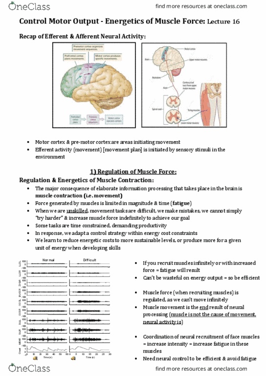
What is Motor Control? (not VI)
• Motor control: information processing related activities carried out by the central
nervous system (CNS) that organises the musculoskeletal system to
create coordinated and skilled movements
The study of movement is the field of motor control growing field in neuroscience
What is the information processing model?
Executive (stimulus identification, response selection, response programming) Effector
(Motor program, spinal cord, muscles)
How are movements controlled?
Overview of this lecture
- lower motor neurons (the neurons that directly innervate muscle)
- different types of muscle
- functional substructure of skeletal muscle
- excitation-contraction coupling (how does an action potential cause a physical
movement)
Looking at what really happens when an action potential reaches the muscle and the
molecular basis of contraction.
muscles
find more resources at oneclass.com
find more resources at oneclass.com

Afferent going towards brain
Efferent going away from brain (effector) exit brain
PNS: 2 functional divisions
• Sensory (afferent) division
– Somatic afferent fibers – carry impulses from skin, skeletal muscles and joints
– Visceral afferent fibers – transmit impulses from visceral organs (body
systems)
• Motor (efferent) division
– Transmits impulses from the CNS to effector organs
Dorsal = back
What is found in the dorsal root ganglion?
Cell bodies of sensory neurons – carrying information from mechanoreceptors in skin.
Dorsal – sensory; Ventral – motor.
Ascending = afferent
Descending = efferent
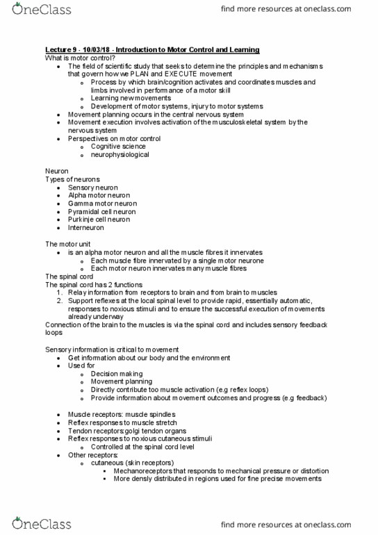
Single alpha motor neuron with cell body in ventral horn. Dendrites cover a large region of
space (and extend vertically up and down the spinal cord. Axon leaves the protection of the
vertebra and targets a muscle
Cell bodies located in the ventral horn of the spinal cord (for control of the body) and in the
motor nuclei of cranial nerves in the brainstem for movements of the eyes, face and
oropharynx.
Axons project out ventral root (mixed with sensory inputs, which enter via dorsal root).
Motor Neurons
Motor neurons reside in the ventral region of the gray matter of the spinal cord.
There is an enlargement of the ventral horn at the cervical region (C3 – T1) as there are a
large number of motor neurons to innervate the skeletal muscle of the upper limbs.
The ventral horn of the lumbar region (L1-L5), which innervates the muscles of the lower
limb, are similarly bigger than the ventral horn of the thoracic region
find more resources at oneclass.com
find more resources at oneclass.com

Lower (alpha) motor neurons
- involved in all movements (voluntary and reflexive)
- directly innervate muscle
- cell bodies in spinal cord
Muscle fibers have two special properties – they can change length; they can generate
force
These motorneurones, which directly innervate the muscles, are often called the lower
motorneurones to distiguish the fro the upper otoreuroes i the rai that
provide input to the spinal cord.
The motor unit and the motor pool
Motor unit =
1 alpha motor neuron + the muscle fibers that it innervates
Small motor units involves <10 muscle fibres (e.g. fingers / eyes)
Large motor units involve >1000 muscle fibres (e.g. calf muscles)
Motor neuron pool =
collection of alpha motor neurons that innervates a single muscle
Motor neurons are clustered in columnar, spinal nuclei called motor neuron pools (or
motor nuclei). All of the motor neurons in a motor neuron pool innervate a single muscle
(Figure 1.4), and all motor neurons that innervate a particular muscle are contained in the
same motor neuron pool. Thus, there is a one-to-one relationship between a muscle and a
motor neuron pool.
Muscle = many muscle fibers
Each individual muscle fiber in a muscle is innervated by one, and only one, motor neuron
(make sure you understand the difference between a muscle and a muscle fiber). A single
motor neuron, however, can innervate many muscle fibers. The combination of an
individual motor neuron and all of the muscle fibers that it innervates is called a motor unit.
The number of fibers innervated by a motor unit is called its innervation ratio.
Ventral horn is nicely organised in
that the medial section innervates
skeletal muscle proximal to the body.
And the lateral section of the ventral
horn innervates the skeletal muscle
distal to the body
find more resources at oneclass.com
find more resources at oneclass.com
Document Summary
1 motor units, muscle types and generating force. At the end of this lecture you should be able to: describe two ways in which force generation is controlled in motor units describe the functional substructure of muscles identify different types of muscle discuss differences in motor units. What is motor control? (not vi: motor control: information processing related activities carried out by the central nervous system (cns) that organises the musculoskeletal system to create coordinated and skilled movements. The study of movement is the field of motor control growing field in neuroscience. Executive (stimulus identification, response selection, response programming) effector (motor program, spinal cord, muscles) Overview of this lecture lower motor neurons (the neurons that directly innervate muscle) Different types of muscle functional substructure of skeletal muscle. Excitation-contraction coupling (how does an action potential cause a physical movement) Looking at what really happens when an action potential reaches the muscle and the molecular basis of contraction.