BMS2011 Lecture Notes - Lecture 19: Gastrocnemius Muscle, Pronator Teres Muscle, Flexor Digitorum Longus Muscle

30 May 2018
School
Department
Course
Professor

Week 10. Limb MSK and Human Evolution 1
LIMB MSK PART 2
• Functional bipeds through massive alterations in muscle belly size, insertion patterns and
fundamental reorganisation of the lower limb appendicular
-> shifted because of functional demands
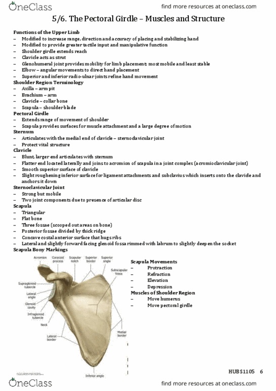
• Pectoral girdle:
o Emphasis on overall mobility
o There is a bony bridge between the upper limb girdle and axial skeleton, sternoclavicular
joint
o Combination of sternoclavicular joint and glenohumeral joint allows full range of
mobility in the upper limbs
• Joints:
Joint
Classification
function
Sternoclavicular joint
Double plane
joint
o Meniscus (pad of fibrous cartilage) exists inside
articular joint -> divides joint in two halves
-> like ball and socket joint
o Increases range of mobility beyond traditional
classic plane joint
o Is the only bridge
o Sternoclavicular ligament allows the bone to
circumduct -> rotate in a circle
o High mobility = sacrificed stability
o Costoclavicular ligament
o Subclavius muscle: very small, extends deep to
clavicle and contracts to stabilise clavivle towards
sternal end
Glenohumeral joint
Ball and socket
joint
o Maximally mobile
o Minimal joint congruence -> head of humerus
ad sapula dot ath er ell
o Glenoid labrum provides stability by increasing
joint congruence
-ridge of fibrocartilage that that encircles glenoid
fossa and increase joint congruence and tendons
of rotator cuff
o Supraspinatus, infraspinatus, teres minor, and
subscapularis muscles enclose the head of the
humerus and help stabilise joint- rotator cuff
o Weak on inferior aspect
Hip joint / pelvic girdle
o Major alterations to hip joint
o Fusion of 3 separate elements
o Femoral head articulating with three bony
elements of oscocca, pubis , illium and ischium
-> forms acetabulum: deep socket that contains
find more resources at oneclass.com
find more resources at oneclass.com

components of all 3 bony elements, good joint
congruence, good match of articular surface of
enlarged head of femur -> more stable ->
transverse ligament increases stability
o Ligament of head of femur: central ligament (not
found in shoulder)
-spans into head of femur
-difference between shoulder and hip joint
o Iliofemoral ligament: along anterior aspect,
limits extension of femur at the hip joint and
prevents hyperextension as we walk
-tight/taunt when femur extended
Elbow joint
o Upper limb
o Mobility
o Pronation: crossing of ulnar over radius to draw
first digit over
o Supination: first digit facing laterally (anatomical
position)
o Critical for abiity to manipulate objects
o Pivot joints proximally and distally
o Proximal and distal radioulnar joint: between
head of radius and the trochloear notch of ulnar
is going to allow pivoting head of radius agants
articular surface
-rounded
-allows pivoting
-held into place by annular ligament: surrounds
head of radius against notch
o Ulnar is stable and in position
(radius = lateral, ulnar = medial)
Knee joint
Hinge-type
synovial joint
o Largest synovial joint
o Fundamentally different joint
o Most complex?
o Specialised modifications in order to take
structures to be both mobile and stable at same
time
o Involves distal femur, proximal tibia and patella
(tibia does not participate in knee joint
articulation – different to elbow joint)
-patella anterior
o Tibia is massively expanded to be weight bearing
bone
o Not a lot of good joint congruence of femur and
proximal tibia
-tibia is flat whereas femur is rounded
-> biomechanics of how we move bipedally
-> we have to shift body weight from lateral to
medial
find more resources at oneclass.com
find more resources at oneclass.com

-> pelvic girdle -> wide -> not mechanically
advantageous
o Carrying angle to femur -> as we plant foot,
rotation to distal femur relative to tibia
-> mismatch in angle (femur has to be at an angle
o Medial and lateral menisci
o Meniscus increases match but are deformable
(softer tissue to match surfaces)
o Tibial (medial)/fibular (lateral) collateral
ligaments
o Intracapsular ligaments (inside joint capsule, not
in elbow): anterior and posterior cruciate
ligament -> cross eachother and directly tie distal
femur to tibia, stabilises joint surface, allows little
rotation but limits as much as possible to
increase stability
-> tendency to break -> blows in lateral aspect ->
unhappy triad (three structures commonly
injured in combination) -> puts medial collateral
ligament under tension -> ligament tears (first
part of triad) -> directly tied to medial meniscus -
> tears off meniscus (second part of triad) ->
third triad (when leg is planted, anterior cruciate
Is at its tightest -> likely to break as well)
-common injury in sports
Distal upper limb joints:
Radiocarpal joint
Midcarpal joint
Carpometacarpal joint
Metacarophalangeal joint
Interphalangeal joints
o When looking at coronal plane through wrist
joints, carpals are largely held by the radius
o Hand is defined by small bones surrounded by
articular surfaces and very complex flat joint
surfaces that allow them to move together
o Carpel tunnel: a fibrous band of tissues
-> flexor renaculum which bind to scaphoid
creates fibrous bridge which deep to it carries 9
tendons from external hand muscles as well as
median nerve
-movements of hand rapidly with pressure placed
on distal portion of wrist -> sliding tendons
against medial nerve -> easy to irritate and cause
to swell -> carpel tunnel syndrome
o Not a lot of fat paddings
Joints of foot :
Many joints
eg. talocrural joint (ankle)
o In contrast to hand has undergone radical
evolution due to bipedalism
o Emphasis on large relatively immobile tarsal
bones and very flat compact metatarsal and
phalanges
o Adduction of first digit: aligning digits against
each other (first digit has taken on weight
bearing function)
find more resources at oneclass.com
find more resources at oneclass.com
Document Summary
Limb msk part 2: functional bipeds through massive alterations in muscle belly size, insertion patterns and fundamental reorganisation of the lower limb appendicular. Ball and socket joint: maximally mobile, minimal joint congruence -> head of humerus function, meniscus (pad of fibrous cartilage) exists inside articular joint -> divides joint in two halves. Increases range of mobility beyond traditional classic plane joint. Iliofemoral ligament: along anterior aspect, limits extension of femur at the hip joint and prevents hyperextension as we walk. Involves distal femur, proximal tibia and patella (tibia does not participate in knee joint articulation different to elbow joint) Patella anterior: tibia is massively expanded to be weight bearing bone, not a lot of good joint congruence of femur and proximal tibia. > we have to shift body weight from lateral to medial. > pelvic girdle -> wide -> not mechanically advantageous: carrying angle to femur -> as we plant foot, rotation to distal femur relative to tibia.