BIOM3002 Lecture Notes - Lecture 2: Simple Squamous Epithelium, Blood Simple, Tunica Media

26 May 2018
School
Department
Course
Professor

HISTOLOGY LECTURE TWO
Epithelia:
• Endothelium
• Lines inside CVS tube
• Basement membrane has vessels [diffusion]
• Avascular
• Innervated
• Polarised [upper and lower surfaces]
• Tight cell to cell contacts
• Rapidly regenerates
CONNECTIVE TISSUE:
• ECM [GS and fibres] and cells
• Supports body in some way
• Great variability
• Collagen fibres common in CVS and elastin and reticular
• GS = GAGs, GPs, PGs
• Collagen fibres = flexible, high tensile strength
• Skin = irregular [different directions force]
• Tendon = aligned fibres = dense regular CT
• Dermis = dense irregular CT
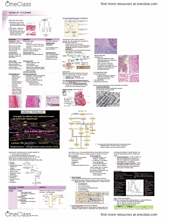
CARDIAC MUSCLE:
• Contract/move
• Synctium [electric] - reacts to ANS and hormones but doesn't
need
• Regular arrangement myofilaments = striated
• Branching fibres
• Single central nucleus
• Many capillaries
• Intercalated discs and gap junctions
SMOOTH MUSCLE: slower, can drive itself, multiple layers right
and loose spiral
TUNICA INTIMA:
• Endothelium = in contact with blood = simple squamous
epithelium
• Sub-endothelium = CT underneath = branches conductive
system
• Protection underlying tissues
• Non-thrombogenic surface
• Regulation tone of media therefore blood flow by production
of vasoactive substances
• Allows diffusion and exchange
• Endocardium in heart
find more resources at oneclass.com
find more resources at oneclass.com

TUNICA MEDIA:
• Myocardium in heart
• Most variable layer
• Determines haemodynamics, BP and blood flow
• Dampens pulsatile flow = smooth flow to organs
• Trabeculae carnae, musculi pectinati and papillary muscles =
extensions
• Large arteries = elastic
• Smaller vessels = smooth muscle
• Absent in capillaries – have isolated SM cells 'pericytes'
TUNICA ADVENTITIA:
• In heart = epicardium = friction-free movement
• Collagen and adipose covered by mesothelium [simple squamous
epithelium]
• Surrounding layer determines interactions with surrounding
structures
• Contains vessels and nerves that supply heart and vessels =
vasa vasorum and nervi vasorum
• In aorta, vasa vasorum = the target of some disease
processes
• In vessels, adventitia adheres to surrounding structures
• Fat = in epicardium and around coronary vessels
CARDIAC MYOCTYES:
• Specialised for conducting – not nervous cells
• Conductive cells pale because no contractile filaments
Elastic artery:
Media > adventitia > intima
Dampens pulsatile flow [distends and recoils] - elastin
Collagen > smooth muscle cells > elastin
Muscular artery:
• Distributors, control blood flow and BP
• Often arteries paralleled with veins 'vena comitans'
• Adventitia = collagen
• Media = SM [controls callibre vessels]
• Intima = elastin [single band = internal elastic lamina;
large arteries may have external elastic lamina]
Arteries vs veins:
A: thick wall relative to lumen; wall maintains circular
shape; tunica media thickest layer; layers are distinct
V: thin wall relative to lumen; wall often collapsed, tunica
adventitia thickest layer; layers, particularly media and
adventitia, not as distinctive; larger veins can have
longitudinal smooth muscle in adventitia
find more resources at oneclass.com
find more resources at oneclass.com