BIOM3002 Lecture Notes - Lecture 6: Distal Convoluted Tubule, Proximal Tubule, Macula Densa

26 May 2018
School
Department
Course
Professor

HISTOLOGY LECTURE SIX
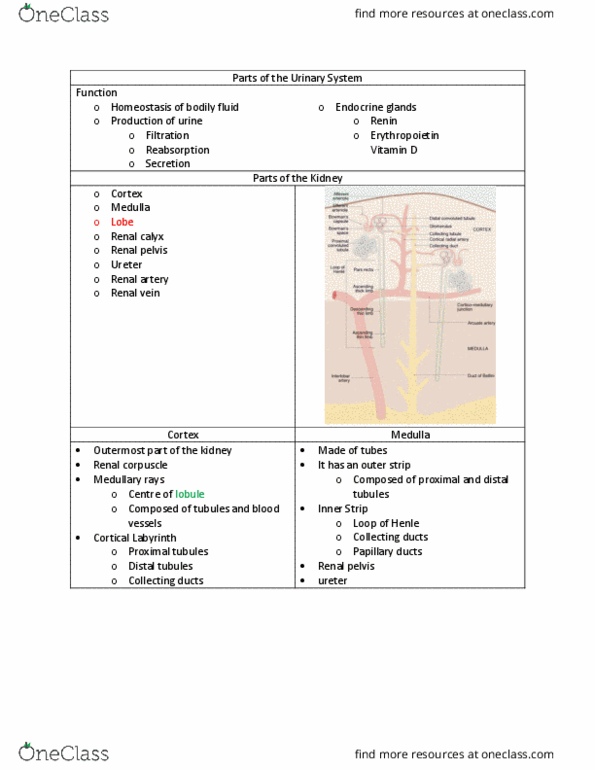
Lobe = medullary pyramid and associated cortical tissues
Lobule = medullary ray and surrounding cortical tissue
If you see renal corpuscles you know you are in: glomeruli;
cortex [arteriole in and out]
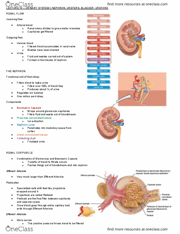
Renal corpuscle: bowman's capsule and glomerulus
• Afferent arteriole supply
• Blood filtered through capillaries
• Single layer fenestrated epithelium
• Blood leaves via efferent arterioles
• Ultrafiltrate -> Bowman's space
• Bowman's capsule = visceral [podocytes] and parietal [simple
epithelium] layers
Renal tubule: continuous with Bowman's capsule
Three methods to increase blood pressure:
1. Renin – vasoconstriction
2. Afferent > efferent arteriole
3. Increasing absorption [Ang I/II + aldosterone] from primary
nephron
At BP < 60: kidney cannot function
Podocytes: interdigitate; form splits; processes – pedicles –
pores [size] allows molecules to pass from glomeruli to
Bowman's capsule
Basement membrane underneath is negatively charged therefore
(-) charged proteins are driven away
Filtration system:
• Capillary endothelium [fenestrated]
• Thick glomerular basement membrane
• Pedicles of podocytes [visceral layer Bowman's capsule] and
filtration slit diaphragm
Vascular pole: afferent and efferent arterioles
Urinary pole: primary urine to proximal convoluted tubule
Other cells in this image = MACULA DENSA: chemoreceptors –
secrete renin [part of thick ascending part – distal
convoluted tubule] [BP restores]
Outstretched cells, look like macula densa but spread-out
[some in glomerulus, some outside]: macrophages/mesangial
cells
find more resources at oneclass.com
find more resources at oneclass.com
Document Summary
Lobe = medullary pyramid and associated cortical tissues. Lobule = medullary ray and surrounding cortical tissue. If you see renal corpuscles you know you are in: glomeruli; cortex [arteriole in and out] Renal corpuscle: bowman"s capsule and glomerulus: afferent arteriole supply, blood filtered through capillaries, single layer fenestrated epithelium, blood leaves via efferent arterioles, ultrafiltrate -> bowman"s space, bowman"s capsule = visceral [podocytes] and parietal [simple epithelium] layers. Three methods to increase blood pressure: renin vasoconstriction, afferent > efferent arteriole, increasing absorption [ang i/ii + aldosterone] from primary nephron. Podocytes: interdigitate; form splits; processes pedicles pores [size] allows molecules to pass from glomeruli to. Basement membrane underneath is negatively charged therefore (-) charged proteins are driven away. Filtration system: capillary endothelium [fenestrated, thick glomerular basement membrane, pedicles of podocytes [visceral layer bowman"s capsule] and filtration slit diaphragm. Urinary pole: primary urine to proximal convoluted tubule.